Type III Hypersensitivity
Write in Short Type 3 hypersensitivity reaction.
Answer:
Type 3 hypersensitivity reaction is also known as an immune complex-mediated reaction or Arthus reaction.
Type 3 reactions result from the deposition of antigen—antibody complexes on tissues, which is followed by activation of the complement system and inflammatory reaction, resulting in cell injury. The onset of type 3 reaction takes place about 6 hours after exposure to the antigen.
Type 3 Hypersensitivity Etiology
There are 3 types of possible etiologic factors precipitating type 3 reaction:
1. Persistence of lowgrade microbial infection: A low-grade infection with bacteria or viruses stimulates a somewhat weak antibody response. The persistence of infection (antigen) and corresponding weak antibody response leads to chronic antigen-antibody complex formation. Since these complexes fail to get eliminated from body fluids, they are instead deposited in tissues, For Example. in blood vessel walls, glo- merely, joint tissue, etc.
2. Extrinsic environmental antigen: Exogenous antigens may be inhaled into the lungs, For Example. antigens derived from molds, plants, or animals. The inhaled antigen combines with antibodies in the alveolar fluid and forms antigen—antibody complex which is deposited in the alveolar walls.
3. Autoimmune process: Another sequence in type 3 reaction can be the formation of autoantibodies against their own tissue (self-antigen) forming an autoantibody-self antigen complex. Such self-antigens can be circulating (For Example. IgA) or tissue derived (For Example. DNA). Immune complexes containing both components from the body’s own system can thus be deposited in tissues.
Type III hypersensitivity examples
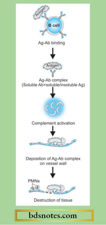
Type 3 hypersensitivity Pathogenesis
Type 3 reaction has participation by IgG and IgM antibodies, neutrophils, mast cells, and complement. The sequence of underlying mechanisms is as follows:
- Immune complexes are formed by the interaction of soluble antibodies and soluble or insoluble antigens.
- Immune complexes which fail to get removed from body fluid get deposited into tissues. Generally, small and intermediate-sized antibodies and antigens precipitate out of the body’s fluid and get deposited in tissues.
- Fc component of antibody links with complement and activates the classical pathway of complement resulting in the formation of C3a, C5a, and membrane attack complex.
- C3a stimulates the release of histamine from mast cells and its resultant effects of increased vascular permeability and edema.
- C5a releases proinflammatory mediators and chemotactic agents for neutrophils.
- Accumulated neutrophils and macrophages in the tissue release cytokines and result in tissue destruction.
Type III hypersensitivity in SLE
Examples of Type 3 Reaction
Common examples of cell injury by type III injury are as under:
- Immune complex glomerulonephritis in which the antigen may be glomerular basement membrane or exogenous agents (For Example. Streptococcal antigen).
- Systemic lupus erythematosus in which there is a nuclear antigen (DNA, RNA) and there is the formation of antinuclear and anti-DNA autoantibodies.
- Rheumatoid arthritis in which there is a nuclear antigen.
- Farmer’s lung in which actinomycetes-contaminated hay acts as an antigen.
- Polyarteritis nodosa and Wegener’s granulomatosis with antineutrophil cytoplasmic antigen.
Leave a Reply