Tubercular Granuloma
Discuss the formation of tubercular granuloma.
Or
Write short note on tubercular granuloma.
Or
Write in brief the granuloma of tuberculosis.
Answer:
Granuloma is a tumor-like proliferation of granulation tissue.
It is seen in granulomatous nodule in rheumatoid arthritis and rheumatic fever.
Tubercular granuloma
Formation of Tubercular Granuloma
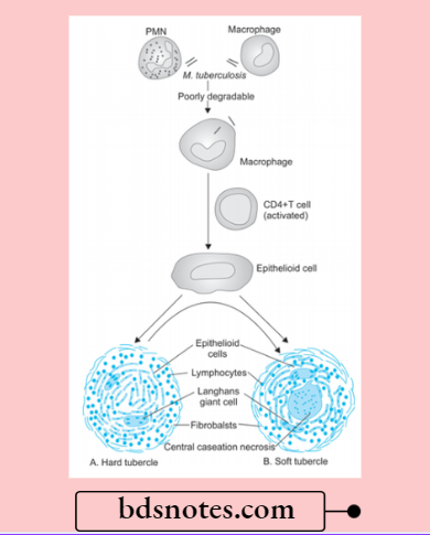
- As the tubercle bacilli are injected intravenously, the bacilli are lodged in pulmonary capillaries where an initial response of neutrophils is evoked which are rapidly destroyed by the organisms. There are two types of cells which are essential for a response to tubercle bacilli, i.e. macrophages and T cells.
- After about l2 hours, there is progressive infiltration by macrophages. This is due to coating of tubercle bacilli with serum complement factors C2a and C3b which act as opsonins and attract the macrophages.
- Macrophages start phagocytosing the tubercle bacilli and either try to kill the bacteria or die away themselves. When macrophages die themselves they produce nitric oxide radicals which have antimycobacterial properties and also Cause increased synthesis of cytokines (TNF-a and IL-1) resulting in the proliferation of macrophages locally as well as increased recruitment from blood monocytes.
- As a part of body’s immune response, T and B cells are activated. Activated CD4+T cells elaborate cytokines, IFN-? and lL-2. These cytokines and their regulators determine the host’s response by infiltrating macrophage monocytes and develop the cell-mediated delayed-type hypersensitivity reaction.
- B cells form antibodies but humoral immunity plays litte role in body’s defense against tubercle bacilli.
- In 2-3 days, the macrophages undergo structural changes as a result of immune mechanisms—the cytoplasm becomes pale and eosinophilic and their nuclei become elongated and vesicular. These modified macrophages resemble epithelial cells and are called epithelioid cells (i.e. epithelial-like).
- Epithelioid cells in time aggregate into tight clusters or granulomas. Release of cytokines in response to sensitized CD4+T cells and some constituents of mycobacterial cell wall play a role in formation of granuloma.
Histology of tuberculous granuloma
- Some macrophages, unable to destroy tubercle bacilli, fuse together and form multinucleated giant cells. These giant cells may be Langhans type having peripherally arranged nuclei in the form of horseshoe or ring, or clustered at the two poles of the giant cell; or they may be foreign body type having centrally placed nuclei.
- Around the mass or cluster of epithelioid cells and a few giant cells, a zone of lymphocytes and plasma cells is formed which is further surrounded by fibroblasts. The lesion at this stage is called hard tubercle due to the absence of central necrosis.
- Within 10-14 days, the center of the cellular mass begins to undergo caseation necrosis, characterized by a cheesy appearance and high lipid content. This stage is called soft tubercle which is the hallmark of tuberculous lesions.
- The soft tubercle which is a fully-developed granuloma with a caseous center does not favor rapid proliferation of tubercle bacilli.
Leave a Reply