Triangles Of The Neck
Describe the boundaries of scalene triangle in brief and enumerate the structures passing through it.
Answer.
Scalene Triangle Location
“Early Signs Of Issues In Triangles Of The Neck”
Root of the neck
“Importance Of Triangles Of The Neck In Medical Practice”
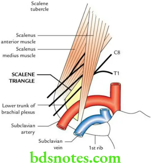
Scalene Triangle Boundaries
Scalene Triangle Anterior:
Scalenus anterior
Scalene Triangle Posterior:
Scalenus medius
“Understanding The Role Of Triangles Of The Neck In Anatomy”
Scalene Triangle Base:
1st rib
Scalene Triangle Apex:
Meeting point of the scalenus anterior and scalenus medius
Scalene Triangle Structures passing through this triangle
- Subclavian artery
- Brachial plexus (lower trunk)
“Step-By-Step Guide To Studying Triangles Of The Neck Effectively”
Scalene Triangle Applied anatomy
- Scalene syndrome: Occurs due to compression of the lower trunk of brachial plexus and subclavian artery in scalene triangle due to (a) spasm of scalene muscles or (b) presence of cervical rib.
- Clinically, it presents as:
- Tingling and numbness in the area of distribution of C8 and T1.
- Progressive wasting of intrinsic muscles of the hand due to involvement of C8 and T1.
- Absence of radial pulse due to compression of the subclavian artery.
“Tips To Prevent Complications From Neck Triangle Issues”
Triangles Of The Neck
Describe various triangles of neck and their boundaries.
Discuss the differential diagnosis of neck swelling.
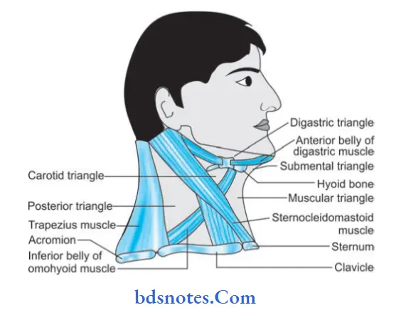
Answer. Each side of neck is the quadrilateral space which is subdivided by sternocleidomastoid into anterior triangle and posterior triangle.
These triangles are further subdivided into
- Anterior Triangle
- Posterior Triangle
“Comprehensive Overview Of Triangles Of The Neck And Their Functions”
“How To Live A Healthier Life While Managing Neck Triangle Health”
Anterior Triangle Boundaries
- Anterior: Anterior midline of the neck extending from symphysis menti above to the middle of suprasternal notch below.
- Posterior: Anterior border of sternocleidomastoid
- Base: Lower border ofthe body ofmandible and line joining the angle of mandible with the mastoid process
- Apex: Suprasternal notch, at the meeting point between anterior border of sternocleidomastoid and anterior midline.
Subdivisions of anterior triangle
The anterior triangle in subdivided by the diagastric muscle and superior belly of omohyoid into following:
- Submental triangle
- Digastric triangle
- Carotid triangle
- Muscular triangle.
“The Role Of Imaging In Diagnosing Triangles Of The Neck Issues Accurately”
Submental triangle
This triangle is complete only when the neck is seen from the front. Each half of the triangle is visible when viewed from side.
Submental triangle Boundaries
- On each side: Anterior belly of diagastric
- Base: Body of hyoid bone
- Apex: Chin or symphysis menti
- Floor: Oral diaphragm formed by the mylohyoid muscles.
Digastric Triangle Boundaries
- Anteroinferior: Anterior belly of digastric
- Posteroinferior: Posterior belly of digastric
- Base: Base of the mandible and an imaginary line joining the angle of mandible to the mastoid process
- Apex: Intermediate tendon of diagastric muscle bound down to hyoid bone by a facial sling.
- Floor: Is formed by mylohyoid muscle (anteriorly), hyoglossus muscle and small partofmiddle constrictor(posteriorly).
- Roof: Is formed by the investing layer of deep cervical fascia which splits to enclose the submandibular salivary gland.
“Best Practices For Treating Triangles Of The Neck Symptoms Safely”
Carotid triangle Boundaries
- Superior: Posterior belly of digastric and stylohyoid
- Anteroinferior: Superior belly of omohyoid
- Posterior: Anterior border of sternocleidomastoid
- Roof: Is formed by investing layer of deep cervical fascia
- Floor: Is formed by four muscles
1. Thyrohyoid
2. Hyoglossus
3. Middle constrictor of pharynx
4. Inferior constrictor of pharynx.
Muscular triangle Boundaries
- Anterior: Anterior midline of the neck
- Antero-superior: Superior belly of the omohyoid
- Posteroinferior: Anterior border of sternocleidomastoid.
Posterior triangle Boundaries
- Anterior: Posterior border of sternocleidomastoid
- Posterior: Anterior border of trapezius
- Base: Middle third of the clavicle
- Apex: Meeting point of sternocleidomastoid and trapezius on the superior nuchal line.
- Roof: Is formed by investing layer of deep cervical fascia stretching between sternomastoid and trapezius muscles
- Floor: Is muscular and is formed by following muscles
“How To Cope With Challenges Of Triangles Of The Neck-Related Issues”
From above downwards
- Semispinalis capitis
- Splenius capitis
- Levator scapulae
- Scalenus posterior
- Scalenus medius
- Outer border of 1st rib.
Sub-divisions of Posterior triangle
- Occipital triangle
- Supraclavicular triangle
Occipital Triangle (From above downwards)
Occipital artery at apex
Spinal part of accessory nerve
Four cutaneous branches of cervical plexus of nerves
1. Lesser occipital
2. Great auricular
3. Transverse cervical
4. Supra clavicular.
Muscular branches of C3 and C4 nerves
Dorsal scapular nerve.
“Why Early Intervention Is Critical For Triangles Of The Neck Outcomes”
Supraclavicular triangle
Trunks of brachial plexus of nerves with their branches
- Dorsal scapular
- Long thoracic
- Nerve to subclavius.
Subclavian artery — 3rd part
Subclavian vein
External jugular vein
Supraclavicular lymph nodes.
Leave a Reply