• Skip to main content
  • Skip to secondary menu
  • Skip to primary sidebar
  • About Us
  • Terms of Use
  • Privacy Policy
  • Disclaimer
  • Contact Us
  • Sitemap

BDS Notes

BDS notes, Question and Answers

  • Public Health Dentistry
  • Periodontics
  • Pharmacology
  • Pathology
  • Orthodontics
    • Anchorage In Orthodontics
    • Mandibular Growth, Functional Matrix
    • Retention and Relapse
  • General Surgery
    • Cysts: Types, Causes, Symptoms
    • Maxillofacial Fractures, Disorders, and Treatments
    • Lymphatic Disorders
    • Neurological and Facial Disorders
  • Temporal And Infratemporal Regions
    • Spinal and Neuroanatomy
  • Dental Materials
    • Dental Amalgam
Home » Treatment Of Gingival Enlargement

Treatment Of Gingival Enlargement

March 3, 2025 by Kristensmith Taylor Leave a Comment

Treatment Of Gingival Enlargement

“What is gingival enlargement and how is it treated?”

Enumeration of Techniques Performed to Increase Width of Attached Gingiva.

  • Gingival augmentation apical to area of recession:
    • Free epithelial autograft
    • Free connective tissue autograft
    • Apically positioned flap
    • Fenestration
    • Vestibular extension.

“Understanding the role of gingival enlargement in oral health”

  • Gingival augmentation coronal to recession or root coverage
    • Free epithelial autograft
    • Free connective tissue autograft
    • Pedicle autografts
      • Rotational, i.e. lateral pedicle flap and double papilla flap
      • Advanced, i.e. Coronally displaced flap and semilunar flap
    • Subepithelial connective tissue graft
    • Subpedicle connective tissue
    • Pouch and tunnel technique
    • Envelope technique
    • Apically displaced flap surgery
    • Guided tissue regeneration technique
      Used for both pocket eradication and widening of zone of attached gingiva:

“Steps to identify common causes of gingival enlargement”

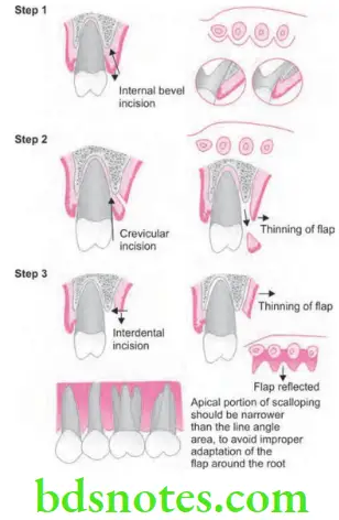
      • Step 1: Internal bevel incision is made 1 mm from the crest of the gingiva and directed towards the crest of the bone
      • Step 2: Crevicular incision is made followed by initial elevation of flap and than interdental incision is performed, the wedge of the tissue containing the pocket wall is removed.
      • Step 3: Vertical releasing incisions are made extending beyond the mucogingival junction and flap is elevated with a periosteal elevator.
      • Step 4: Remove all the granulation tissues, root planning is done and flap is positioned apically at the tooth bone junction.
      • Step 5: Flaps are sutured together.

“Importance of studying gingival enlargement for better dental care”

Periodontics Mucogingival Surgery Periodontal Plastic Surgery Apically displaced flap surgery

Filed Under: Periodontics

Reader Interactions

Leave a Reply Cancel reply

Your email address will not be published. Required fields are marked *

Primary Sidebar

Recent Posts

  • Branchial Cleft Cyst: Background, Pathophysiology, Etiology
  • Maxillary Nerve: Origin, Course, And Branches
  • The Father Of Anatomy And A Great Anatomist Herophilus
  • Bone Structure – Anatomy
  • The External Carotid Artery: Anatomy, Branches, And Functions
  • Occipitofrontalis Muscle
  • Superficial Temporal Artery
  • Platysma Muscle
  • Cartilage
  • Cauda Equina And Conus Medullaris Syndromes
  • Subcutaneous Injections And Device Management
  • Types Of Circulation: Pulmonary, Systemic, And Portal
  • Hierarchical Organization Of Skeletal Muscle Tissue
  • Elastic Cartilage Histology Short Note For Medical Exams
  • Cellular Organelles And Structure
  • The Golgi Apparatus – The Cell
  • The Cytoplasmic Inclusions Of Certain Plant Cells
  • Dental Abscess
  • Laser Surgery
  • Our Facial Muscles And Their Functions

Copyright © 2026 · Magazine Pro on Genesis Framework · WordPress · Log in