• Skip to main content
  • Skip to secondary menu
  • Skip to primary sidebar
  • About Us
  • Terms of Use
  • Privacy Policy
  • Disclaimer
  • Contact Us
  • Sitemap

BDS Notes

BDS notes, Question and Answers

  • Public Health Dentistry
  • Periodontics
  • Pharmacology
  • Pathology
  • Orthodontics
    • Anchorage In Orthodontics
    • Mandibular Growth, Functional Matrix
    • Retention and Relapse
  • General Surgery
    • Cysts: Types, Causes, Symptoms
    • Maxillofacial Fractures, Disorders, and Treatments
    • Lymphatic Disorders
    • Neurological and Facial Disorders
  • Temporal And Infratemporal Regions
    • Spinal and Neuroanatomy
  • Dental Materials
    • Dental Amalgam
Home » Transseptal Fibers: The Key To Tooth Stability And Orthodontic Relapse

Transseptal Fibers: The Key To Tooth Stability And Orthodontic Relapse

February 5, 2026 by Kristensmith Taylor Leave a Comment

Transseptal Fibers: The Key To Tooth Stability And Orthodontic Relapse

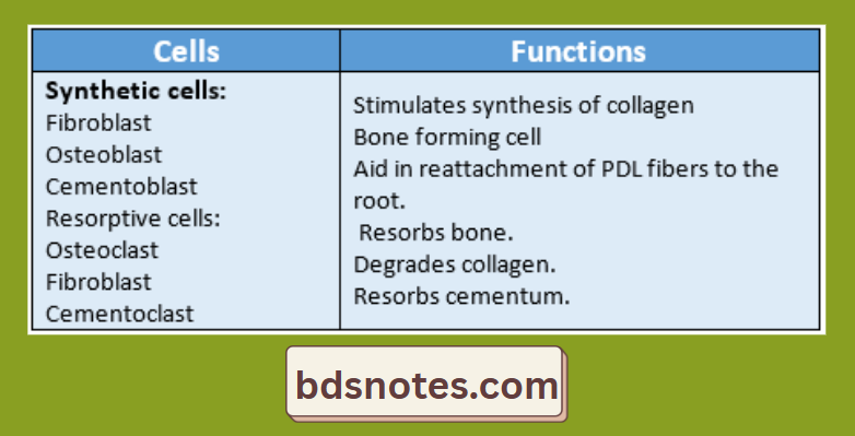
Question 1. Synthetic cells of PDL.
Answer:

Periodontal Tissue Remodeling Cells

Question 2. Transseptal fibers.
Answer:

  • These fibers run interdentally from the cementum apical to the junctional epithelium of one tooth over the alveolar crest to a similar region of the adjacent tooth.
  • By these, all tire teeth are connected in an arch.
  • They are responsible for post-retention relapse of orthodontic treatment.
  • They are capable of turnover and remodeling under normal physiologic conditions and therapeutic tooth movement.
  • They ensure clinical stability of tooth position.

Question 3. Bundle fibers of the periodontal membrane.
Answer:

Fiber bundles composing ligament:

1. Dentogingival group:

  • Extends from cervical cementum to lamina propria of the free and attached gingiva.

2. Alveologingival group:

  • Extends from the bone of the alveolar crest to lamina propria of the free and attached gingiva.

3. Circular group:

  • It forms a band around the neck of the tooth.

4. Dentoperiosteal group:

  • Runs apically from the cementum up to the alveolar process.

5. Transseptal fiber:

  • Run interdentally from the cementum of one tooth to the cementum of the adjacent tooth.

Question 4. Age changes in the periodontal ligament.
Answer:

  • The cell number and activity decrease with age
  • PDL fibers become attached to the scalloping ends of the alveolar bone.
  • PDL activity decreases.
  • Destructive changes occur due to the presence of gingival and periodontal diseases in old age.
  • Some of the teeth become non-functional.
  • PDL width decreases.

Filed Under: Anatomy

Reader Interactions

Leave a Reply Cancel reply

Your email address will not be published. Required fields are marked *

Primary Sidebar

Recent Posts

  • Branchial Cleft Cyst: Background, Pathophysiology, Etiology
  • Maxillary Nerve: Origin, Course, And Branches
  • The Father Of Anatomy And A Great Anatomist Herophilus
  • Bone Structure – Anatomy
  • The External Carotid Artery: Anatomy, Branches, And Functions
  • Occipitofrontalis Muscle
  • Superficial Temporal Artery
  • Platysma Muscle
  • Cartilage
  • Cauda Equina And Conus Medullaris Syndromes
  • Subcutaneous Injections And Device Management
  • Types Of Circulation: Pulmonary, Systemic, And Portal
  • Structure Of Skeletal Muscle
  • Elastic Cartilage
  • Cellular Organelles And Structure
  • The Golgi Apparatus – The Cell
  • The Cytoplasmic Inclusions Of Certain Plant Cells
  • Dental Abscess
  • Laser Surgery
  • Our Facial Muscles And Their Functions

Copyright © 2026 · Magazine Pro on Genesis Framework · WordPress · Log in