Tooth Pulp Explained: Structure, Cells, And Functions
Describe briefly pulp.
Answer:
Pulp Definition:
The dental pulp is defined as the richly vascularised and innervated connective tissue of mesodermal origin enclosed by dentin with communications to the periodontal ligament.
Functions:
1. Formative:
- It produces the dentin that surrounds it.
2. Nutritive:
- It nourishes the avascular dentin.
3. Protective:
- It carries nerves that give dentin its sensitivity.
4. Reparative:
- It is capable of producing new dentin when required.
5. Inductive:
- Pulp interacts with oral epithelium cells and causes differentiation of dental lamina and results in enamel organ formation which determines the type of tooth.
Anatomy:
- The dental pulp occupies the center of each tooth and consists of soft connective tissue.
- The pulp present in the crown is called coronal pulp and the pulp present in the root is called radicular pulp.
- The pulp consists of apical foramen and accessory canals.
- The apical opening is found on the lateral side of the apex.
- Frequently, there are two or more foramina separated by a portion of dentin and cementum.
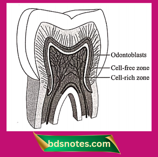
Structure:
- Four distinct zones can be distinguished in the pulp. They are.
- Odontoblastic zone: At the pulp periphery.
- Cell-free zone of Weil:
- Beneath the odontoblasts.
- It is prominent in the coronal pulp.
- Cell-rich zone:
- Adjacent to the cell-free zone.
- Cell density is high in this zone.
- Pulp core:
- It is characterized by the major vessels and nerves of the pulp.
Cellular elements:
1. Odontoblasts:
- They form a layer lining the periphery of the pulp and have a process extending into dentin.
- They are columnar cells with basal nuclei.
- They synthesize and secrete collagen from dentin.
2. Fibroblasts:
- They are the most prominent cells of pulp.
- They are stellate shape cells with extensive processes
- They form and maintain the pulp matrix.
- They are capable of ingesting and degrading collagen.
3. Undifferential ectomesenchyme cells:
- Depending on the stimulus, these cells may give rise to odontoblasts, fibroblasts, or macrophages.
4. Defence cells:
- Histiocytes or macrophages
- Mast cells
- Plasma cells.
5. Other cells:
- Eosinophils and lymphocytes.
- Dendritic cells
Matrix and ground substance:
- Matrix consists of collagen fibers and ground substance composed of principally glycosaminoglycans, glycoproteins, and water.
Blood vessels:
- The pulp is supplied by superior alveolar and inferior alveolar vessels.
Lymph vessels:
- The lymph vessels of anterior teeth drain into submental lymph nodes and that of posterior teeth into submandibular and deep cervical lymph nodes.
Leave a Reply