Tissue Response To Orthodontic Forces
Describe in detail tissue changes in response to mild and heavy orthodontic force.
Or
Describe the histological changes occurring in PDL and alveolar bone when a mild orthodontic force is applied to a tooth, what would happen if a force applied turned into heavy force?
Answer. When force is applied to a tooth during orthodontic treatment it leads to the formation of areas of pressure and tension around the teeth
“Understanding the role of tissue response in orthodontic treatment: Q&A explained”
- Areas of pressure are formed in the direction of tooth movement.
- Areas of tension form in the opposite direction of tooth movement.
- Bone surfaces subjected to tension shows bone deposition.
- The bone surfaces subjected to pressure show bone resorption.
- The histological changes associated with tooth movements depend on the amount and duration of force.
- The histological changes associated with tooth movements are studied under the following headings.
- Changes following application of mild force.
- Changes following application of extreme force.
“Importance of studying tissue response for better orthodontic outcomes: Questions explained”

“Common challenges in analyzing tissue response to orthodontic forces: FAQs provided”
Frontal Resorption In Orthodontics
Changes Following Application of Mild/Light Force
Secondary Remodeling Changes
Bony changes elsewhere (other than the area of tension and pressure) to maintain the width of the alveolar bone.
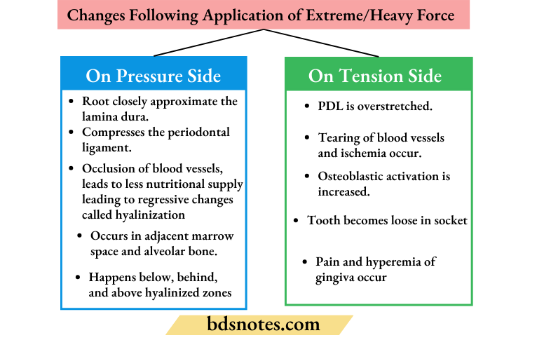
Changes Following Application of Extreme/Heavy Force
On Pressure Side
- Root closely approximates the lamina dura.
- Compresses the periodontal ligament.
- Occlusion of blood vessels leads to less nutritional supply leading to regressive changes called hyalinization.
- Bone resorption occur in the adjacent marrow space and in the alveolar bone, below, behind and above the hyalinized zones this kind of resorption is called undermining or rearward resorption.
“Factors influencing tissue response to orthodontic forces: Q&A”
On Tension Side
- PDL is overstretched.
- Tearing of blood vessels and ischemia occur.
- Osteoblastic activation is increased.
- The tooth becomes loose in the socket.
- Pain and hyperemia of the gingiva occur.
Leave a Reply