Thyroid Gland Development And Anomalies: From Embryo To Adult
Question 1. Development of thyroid gland and its anomalies.
Answer:
Development Thyroid Gland :
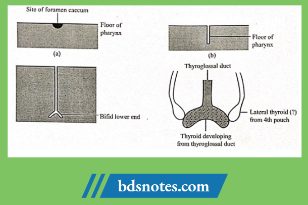
- The thyroid gland develops mainly from the thyroglossal duct.
- The medial ends of the two mandibular arches are separated by a midline swelling called the tuberculum impar.
- Behind the tuberculum, the epithelium is thickened which soon gets depressed to form a diverticulum called thyroglossal duct
- The diverticulum grows down in the midline into the neck.
- Its tip soon bifurcates.
- Proliferation of the cells of the bifid end gives rise to the two lobes of the thyroid gland.
- Parafollicular cells are derived from the caudal pharyngeal complex which is derived from the fourth and fifth pharyngeal pouches.
Thyroid Gland Anomalies:
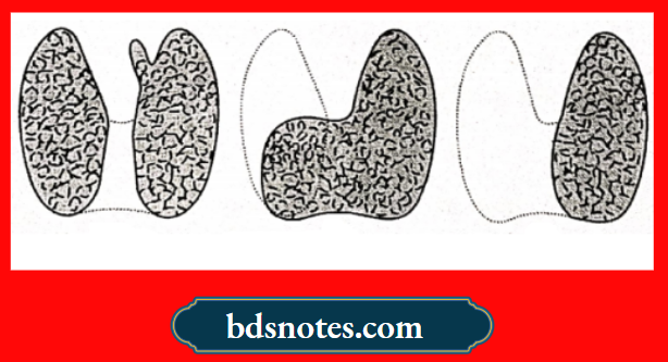
1. Anomalies of shape.
- Pyramidal lobe arises from the isthmus or from one of the thyroid lobes.
- Isthmus may be absent.
- One of the lobes may be small or absent.
2. Anomalies of Position.
- Lingual thyroid – under tongue mucosa.
- Intra-lingual thyroid – embedded in tongue musculature.
- Suprahyoid thyroid – above the hyoid bone.
- Infrahyoid thyroid – below the hyoid bone.
- Intrathoracic thyroid – lie in the thorax.
3. Ectopic Thyroid Tissue.
- Thyroid tissue has been observed in the larynx, trachea, oesophagns, pons, pleura, pericardium and ovaries.
4. Remnants of the Thryoglassal Duct.
- Thyroglossal cyst- occur along the course of the duct.
- Thyroglossal fistula – opening at foramen caecum.
- Carcinoma of the thyroglossal duct.
Question 2. Development of Mandible.
Answer:
- About 4th week of intrauterine life, pharyngeal arches are laid down on the lateral and ventral aspects of foregut.
- Mandible is developed from first pharyngeal arch is called mandibular arch.
- The mandibular processes of both sides grow towards each other and fuse in the midline.
- This results in formation of lower lip and lower jaw.
Development of Condylar Process:
- It occurs of 5thwek of intrauterine life.
- Mesenchymal condensation occurs above the ventral side of developing mandible.
- Cartilage is formed at 10th week which undergoes ossification to form condylar process.
Development of Coronoid Process:
- At 10-14 week of interauterine life, secondary accessory cartilages.
- This gets incorporated into expanding ramus and forms coronoid process.
Development of Mental Region:
- Ossification of 1 or 2 small cartilages occurs on either side of symphysis.
- It forms the mental region.
Leave a Reply