• Skip to main content
  • Skip to secondary menu
  • Skip to primary sidebar
  • About Us
  • Terms of Use
  • Privacy Policy
  • Disclaimer
  • Contact Us
  • Sitemap

BDS Notes

BDS notes, Question and Answers

  • Public Health Dentistry
  • Periodontics
  • Pharmacology
  • Pathology
  • Orthodontics
    • Anchorage In Orthodontics
    • Mandibular Growth, Functional Matrix
    • Retention and Relapse
  • General Surgery
    • Cysts: Types, Causes, Symptoms
    • Maxillofacial Fractures, Disorders, and Treatments
    • Lymphatic Disorders
    • Neurological and Facial Disorders
  • Temporal And Infratemporal Regions
    • Spinal and Neuroanatomy
  • Dental Materials
    • Dental Amalgam
Home » The Scalene Muscles

The Scalene Muscles

July 26, 2025 by Kristensmith Taylor Leave a Comment

The Scalene Muscles

Question 1. Give origin, insertion & superficial relations of scalenus anterior muscle
Answer:

Scalenus Anterior Muscle:

  • It is a paravertebral muscle

Origin:

  • Anterior tubercles of transverse processes of cervical vertebrae 3, 4, 5 & 6

Insertion:

  • Scalenus tubercle & adjoining ridge on the superior surface of first rib
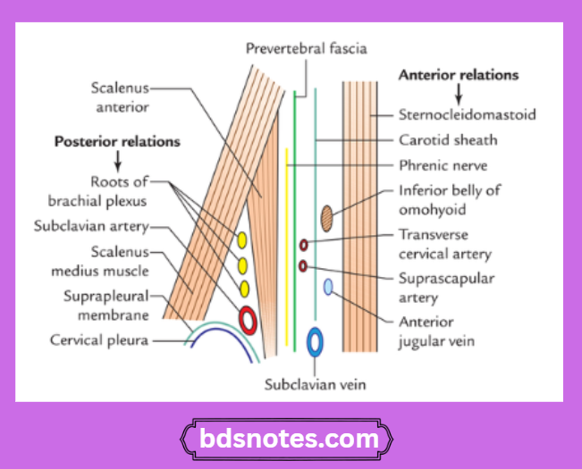
Relations:

“Understanding the scalene muscles through FAQs”

Scalenus Anterior Relations Diagram

“Importance of studying the scalene muscles for medical students: Questions explained”

Sagittal Section Scalenus Anterior Anatomy

Question 2. Thyroid gland follicle
Answer:

  • Thyroid gland is covered by a fibrous capsule
  • Septa extends from capsule into gland substance & divides it into lobules
  • Each lobule is made up of follicles
  • Follicle has a cavity filled with colloid
  • Each follicle consists of

“Common challenges in mastering scalene muscle notes effectively: FAQs provided”

1. Cells:

  1. Follicular cells
    • They line the follicles
    • They secrete T3 & T4 hormones
    • They contains
      • Golgi complex
      • Lysosomes
      • Microtubules
      • Microfilaments
      • Secretory vacuoles
        • They vary in shape according to their activity
          1. NormallyCells are cuboidal with moderate colloid
          2. Inactive cellare flat with abundant colloid
          3. Highly active cellare columnar with scanty colloid
  2. C cells or parafollicular cells
    • They are polyhedral cells with oval nuclei

“Steps to explain cell types involved in the scalene muscles: Myoblasts vs fibroblasts: Q&A guide”

Location:

  • In between follicular cells & basement membrane
  • In connective tissue between the follicles

Contains:

  • Granular endoplasmic reticulum
  • Golgi complex
  • Mitochondria
  • Membrane bound Secretory granules

“Factors influencing success with scalene muscle studies: Q&A”

Hormone secreted:

  • Thyrocalcitonin

2. Connective tissue stroma:

  • It surrounds the follicles
  • It contains capillary plexus, lymphatic capillaries & sympathetic nerves

Filed Under: Anatomy

Reader Interactions

Leave a Reply Cancel reply

Your email address will not be published. Required fields are marked *

Primary Sidebar

Recent Posts

  • Branchial Cleft Cyst: Background, Pathophysiology, Etiology
  • Maxillary Nerve: Origin, Course, And Branches
  • The Father Of Anatomy And A Great Anatomist Herophilus
  • Bone Structure – Anatomy
  • The External Carotid Artery: Anatomy, Branches, And Functions
  • Occipitofrontalis Muscle
  • Superficial Temporal Artery
  • Platysma Muscle
  • Cartilage
  • Cauda Equina And Conus Medullaris Syndromes
  • Subcutaneous Injections And Device Management
  • Types Of Circulation: Pulmonary, Systemic, And Portal
  • Structure Of Skeletal Muscle
  • Elastic Cartilage
  • Cellular Organelles And Structure
  • The Golgi Apparatus – The Cell
  • The Cytoplasmic Inclusions Of Certain Plant Cells
  • Dental Abscess
  • Laser Surgery
  • Our Facial Muscles And Their Functions

Copyright © 2026 · Magazine Pro on Genesis Framework · WordPress · Log in