The Axillary Region
Question 1. Enumerate the structures piercing the clavipectoral fascia.
Answer.
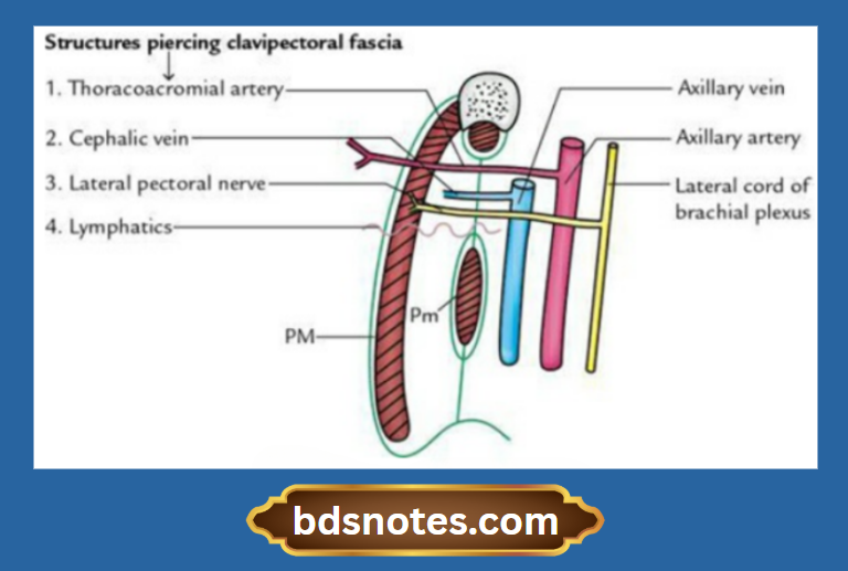
Clavipectoral fascia is pierced by four structures:
- Lateral pectoral nerve
- Thoracoacromial artery
- Cephalic vein
- Lymphatics from the infraclavicular nodes and the deep part of the breast to an apical group of axillary lymph nodes
Axilla
The axilla is a pyramid-shaped space between the upper part of the arm and the thorax.
axillary region
Question 1. Describe the axilla under the following headings: (a) boundaries, (b) contents and (c) applied anatomy.
Answer.
Axilla Boundaries
Axilla Anterior wall:
Axilla Anterior wall is formed by:
- Pectoralis major
- Subclavius muscle
- Clavipectoral fascia
- Pectoralis minor
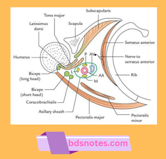
Posterior wall:
The posterior wall is formed by:
- Latissimus dorsi
- Teres major
- Subscapularis
Medial wall:
The medial wall is formed by the serratus anterior muscle, covering the upper part of the lateral thoracic wall (upper 4–5 ribs).
Lateral wall:
The lateral wall is narrow and formed by the intertubercular sulcus of the shaft of the humerus, which contains coracobrachialis and the short head of the biceps brachii.
axilla anatomy
Apex (also called cervicoaxillary canal):
The cervicoaxillary canal is triangular and directed upwards and medially towards the root of the neck. It is bounded:
- Anteriorly, by the posterior border of the clavicle
- Medially, by the outer border of the 1st rib
- Posteriorly, by the upper border of the scapula
Base:
The base is formed by the axillary fascia extending between the anterior and posterior axillary folds.
Base Contents
- Axillary artery and its branches
- Axillary vein and its tributaries
- Cords of the brachial plexus
- Axillary lymph nodes
- Fibrofatty tissue
- Long thoracic and intercostobrachial nerves
- Axillary tail of breast (tail of Spence)
Base Applied anatomy
Axillary abscess:
It occurs due to infection and suppuration of the axillary lymph nodes. Axillary abscess is drained by giving an incision midway between the anterior and posterior axillary folds. The direction of the edge of the knife should face towards the medial wall.
Lymphadenopathy:
Axillary lymph nodes are often infected and enlarged. They should be removed very carefully because of their relationship to major vessels.
Boils:
Due to the presence of abundant hair follicles in axilla, the infection of hair follicles and sebaceous glands is very common and gives rise to multiple boils in the axilla.
Axillary pulse:
It can be felt against the lower part of the lateral wall of the axilla.
Leave a Reply