Tentorium Cerebelli
Question 1. Tentorium cerebella
Answer:
- It is tent shaped fold of duramater forming the roof of the posterior cranial fossa
- It separates cerebellum from the occipital lobes of the cerebrum
- It divides cranial cavity into supratentorial & infratentorial compartments
Margins:
- Anterior free margin
- Outer attached margin
Surfaces:
- Superior convex surface
- Inferior concave surface
“Steps to explain cell types involved in the tentorium cerebelli: Fibroblasts vs endothelial cells: Q&A guide”
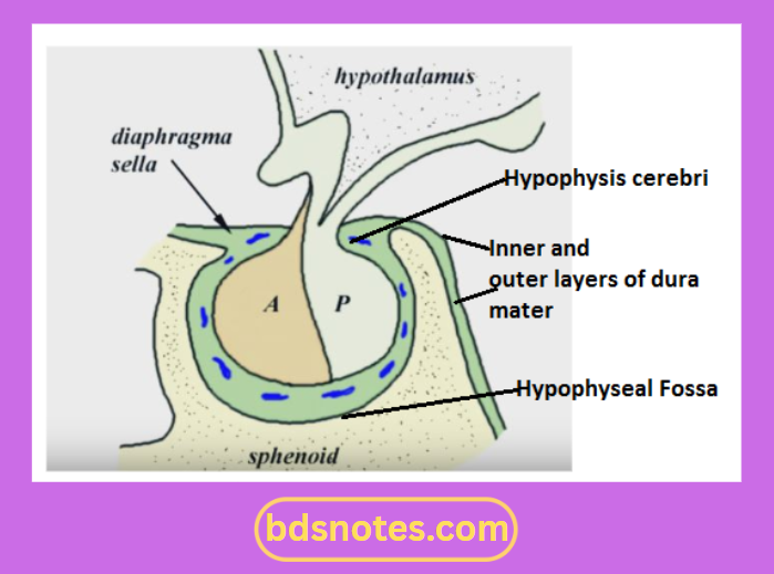
Question 2. Diaphragm sellae
Answer:
- It is a small circular, horizontal fold of duramater forming the roof of the hypophyseal fossa
- It has a central aperture through which the stalk of the hypophysis cerebri passes
Attachments of tentorium cerebelli
Attachments:
1. Anteriorly
- To the tuberculum sellae
2. Posteriorly
- To the dorsum sellae
3. On each side
“Role of fibroblasts in maintaining structural integrity: Questions answered”
- Continuous with the duramater of the middle cranial fossa
“Early warning signs of undiagnosed cell type-related issues: Common questions”
Question 3. Hypoglossal nerve
Answer:
- It is the twelfth cranial nerve
- It supplies the muscles of the tongue
Functional components:
- General somatic efferent
- General somatic afferent
Nucleus:
- It lies in the floor of fourth ventricle beneath Hypoglossal triangle
Branches:
1. Branches containing fibres of the Hypoglossal nerve proper
- They supply extrinsic & intrinsic muscles of the tongue
“Asymptomatic vs symptomatic effects of delayed interventions: Answered”
2. Branches of the hypoglossal nerve containing fibres of nerve C1
- Meningeal branch
- Supplies bone & meninges in the anterior part of the posterior cranial fossa
- Descending branch
- Continues as upper root of the ansa cervicalis
- Branches to the thyrohyoid & geniohyoid muscles
Leave a Reply