Technique For Maxillary Cross-Sectional Occlusal Radiograph
Write a short note on the technique for maxillary cross-sectional occlusal radiograph.
Answer.
Technique for Maxillary Cross-Sectional Occlusal Radiograph
- Image field: This projection shows the palate, the zygomatic process of the maxilla, the anterior-inferior aspects of each antrum, nasolacrimal canals, teeth from the right second molar to the left second molar, and the nasal septum.
- Film placement: The film is placed crosswise into the mouth and gently pushed back until it contacts the anterior border of the rami.
“Understanding the role of maxillary cross-sectional occlusal radiographs in dentistry: Q&A explained”
Maxillary occlusal radiograph
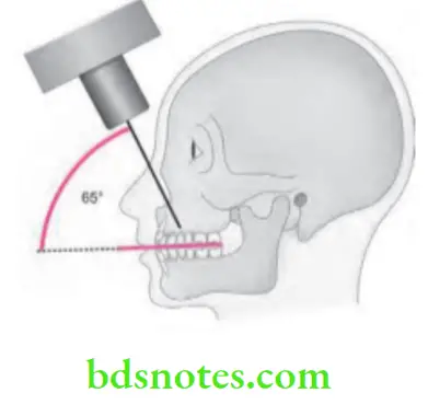
- Projection of the central ray: The central ray is directed at a vertical angulation of +65° and a horizontal angulation of +65° towards the middle of the film. In general, the central ray enters the patient’s face through the Bridge of the nose.
“Common challenges in performing maxillary cross-sectional occlusal radiographs effectively: FAQs provided”
“Importance of studying maxillary cross-sectional occlusal radiograph techniques: Questions explained”
Maxillary occlusal technique
Leave a Reply