Systemic Lupus Erythematosus (SLE): A Multisystem Autoimmune Disorder
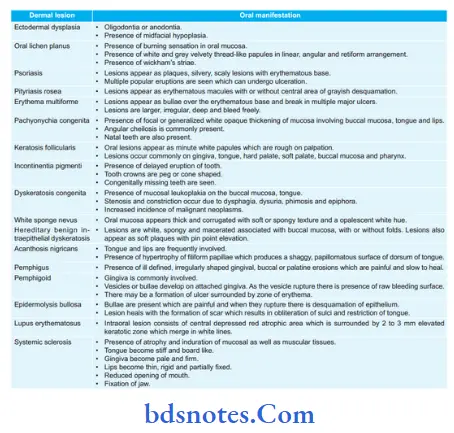
Question. Enumerate dermal lesions with oral manifestations.
Describe systemic lupus erythematosus in detail.
Or
Enumerate the dermal diseases with manifestations.
Describe the etiology, clinical, and histopathological features of systemic lupus erythematosus in detail with a diagram. Give its treatment plan.
“Understanding systemic lupus erythematosus through FAQs: Q&A explained”
Answer. Enumeration of dermal lesions with oral manifestations
“Importance of studying SLE for better diagnostic outcomes: Questions explained”
Systemic lupus erythematosus
It is an autoimmune disease which is characterized by autoantibodies, immune complex formation as well as dysregulation of immune system which causes damage to any organ of body.
Etiology Of Lupus Erythematosus
- Genetic predisposition: A Higher incidence of autoantibodies is seen in blood relatives of the patient.
- Viral infection
- Hormones, i.e., increase estrogen level in pregnancy.
- Autoimmune: Antibodies are developed towards one’s body cells.
“Common challenges in diagnosing systemic lupus erythematosus effectively: FAQs provided”
Pathogenesis Of Lupus Erythematosus
Antibodies are produced in reaction to exposure to normally unexposed self-antigens.
Dysregulation of the immune system leads to excessive production of antibodies against DNA, ribosomes, other nuclear antigens, platelets, erythrocytes, leucocytes, and various tissue-specific antigens, which causes tissue damage.
Clinical Features Of Lupus Erythematosus
- It occurs at 30 years of age in females and 40 years of age in males.
- Female predilection is seen. The female-to-male ratio is 2:1
- The most common sites affected are the face, neck, upper arm, and shoulders. Disease is characterized by repeated remissions and exacerbations over these sites. The patient complained of pain and fever in joints and muscles.
- Itching or burning sensation is also present along with the areas of hyperpigmentation. Symptoms aggravate under exposure to sunlight.
- The characteristic sign of the disease is presence of erythematous patches over the face which coalesce to form roughly symmetrical pattern over the cheeks and across the bridge of nose, this is known as butterfly distribution. In the kidney fibrinoid thickening of glomerular capillaries produces characteristic wire loops, this leads to renal insufficiency.
- In the heart, there isa presence of typical endocarditis involving valves along with fibrinoid degeneration of the epicardium and myocardium.
“Steps to explain causes of systemic lupus erythematosus: Autoimmunity vs genetic predisposition: Q&A guide”
Oral Manifestations Of Lupus Erythematosus
- Buccal mucosa, lip, and palate are most commonly The patient complains of a burning sensation in the mouth. Xerostomia is also seen.
- Lesions have very much similarity to lesions of discoid lupus except that they are hyperemic, edematous and extension of lesion is pronounced. Tendency for bleeding and petechiae is more as well as superfiial ulcerations surrounded by red halo are also present.
- Intraoral lesion consists of a central depressed red atrophic ar,ea which is surrounded by a 2 to 3 mm elevated keratotic zothatich merges with white lines.
Histopathology Of Lupus Erythematosus
- In systemic lupus erythematosus, areas of epithelial atrophy are present with the absence of keratinization.
- There is a presence of liquefactive degeneration of the basal cell layer.
- There is the presence of edema of subepithelial connective tissue with dilatation of vessels.
- In systemic lupus erythematosus, degenerative areas and collagen disturbances are more prominent.
- Inflammatory features are less common.
“Role of immune dysregulation in causing systemic lupus erythematosus: Questions answered”
Laboratory Findings Of Lupus Erythematosus
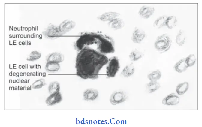
- LE cell inclusion phenomenon is used in which there is addition of blood serum from a person who is suspected to the buff coat of normal blood.
If patient is suffering from systemic lupus erythematosus, typical LE cells will appear. The test consists of rosett of neutrophils surrounding pale nuclear mass. - There is also presence of anemia, leucopenia, thrombocytopenia and elevated ESR and serum gamma globulin level with positive Coombs test.
- Lupus band test is positive, i.e. there is deposition of IgG, IgM or complement component at epidermal dermal junction or basement membrane zone of skin.
“Asymptomatic vs symptomatic effects of ignoring SLE triggers: Q&A”
“Early warning signs of issues addressed by understanding SLE pathogenesis: Common questions”
Treatment Of Lupus Erythematosus
- Exposure to sunlight should be avoided.
- The patient would be kept on systemic corticosteroid therapy.
- NSAIDs should also be given to combat the symptoms.
Leave a Reply