Syphilitic Gumma
Write a short note on syphilitic gumma.
Answer:
Syphilitic gumma is the lesion of tertiary syphilis. Syphilitic gummas are white-gray and rubbery, occur singly or multiply, and vary in size from microscopic defects resembling tubercles to large tumor-like masses.
- They occur in most organs but particularly in skin, subcutaneous tissue, bone, and joints.
- In the liver, scarring of hepatic parenchyma as a result of gummas may cause a distinctive hepatic lesion known as hepatic location.
Syphilitic gumma
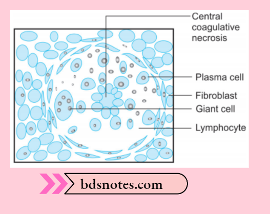
Syphilitic gumma histologically
On histologic examination, the gummas contain a center of coagulated, necrotic material and margins composed of plump or palisaded macrophages and fibroblasts surrounded by large numbers of mononuclear leukocytes, chiefly plasma cells.
Treponemes are scant in this gumma and are difficult to demonstrate.
Gumma in tertiary syphilis
Syphilitic gumma In oral cavity
Oral manifestations of syphilis
- Gumma can occur anywhere in the oral cavity but the more frequent sites are palate, mandible and tongue.
- It occur as a solitary, deep, punched-out ulcer.
- In gumma breathing and swallowing difficulties are encountered by the patients.
- At times perforation of the palatal wall is present.
- Numerous small healed gumma in the tongue result in series of nodules or sparse in the deeper area giving the tongue an upholstered or tufted appearance.
Leave a Reply