• Skip to main content
  • Skip to secondary menu
  • Skip to primary sidebar
  • About Us
  • Terms of Use
  • Privacy Policy
  • Disclaimer
  • Contact Us
  • Sitemap

BDS Notes

BDS notes, Question and Answers

  • Public Health Dentistry
  • Periodontics
  • Pharmacology
  • Pathology
  • Orthodontics
    • Anchorage In Orthodontics
    • Mandibular Growth, Functional Matrix
    • Retention and Relapse
  • General Surgery
    • Cysts: Types, Causes, Symptoms
    • Maxillofacial Fractures, Disorders, and Treatments
    • Lymphatic Disorders
    • Neurological and Facial Disorders
  • Temporal And Infratemporal Regions
    • Spinal and Neuroanatomy
  • Dental Materials
    • Dental Amalgam
Home » Suboccipital Triangle and Suboccipital Muscles

Suboccipital Triangle and Suboccipital Muscles

March 25, 2025 by Kristensmith Taylor Leave a Comment

Suboccipital Triangle and Suboccipital Muscles

Mention boundaries of suboccipital triangle. Describe the contents of the triangle (or) Suboccipital triangle (or) Suboccipital muscles (or) Contents of suboccipital triangle
Answer:

Suboccipital Triangle:

Suboccipital Triangle Boundaries:

“Understanding The Anatomy Of The Suboccipital Triangle”

1. Superomedially:

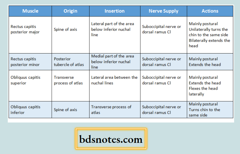
  • Rectus capitis posterior major
  • Rectus capitis posterior minor

2. Superolaterally:

  • Superior oblique muscle

“Anatomy Of The Suboccipital Triangle”

3. Inferiorly:

  • Inferior oblique muscle

Suboccipital Triangle Roof:

  • Medially-semispinalis capitis
  • Laterally-longissimus capitis & splenius capitis

“Role Of The Suboccipital Triangle In Neck Pain”

Suboccipital Triangle Floor:

  • Posterior arch of atlas
  • Posterior atlanto-occipital membrane

Suboccipital Triangle Contents:

1. Third part of vertebral artery:

  • It appers at the foramen transverium of the atlas, grooves the atlas & leaves the triangle by passing deep to the lateral edge of the posterior atlanto-occipital membrane
  • It gives muscular branches to the muscle of the suboccipital region

Suboccipital Triangle and Suboccipital Muscles

“Boundaries Of The Suboccipital Triangle”

2. Dorsal rami of C1 nerve:

  • It emerges between posterior arch of the atlas & the vertebral artery
  • Structures supplied by it are
    • Four suboccipital muscles
    • Semispinalis capitis
    • Nerve to inferior oblique gives off communicating branch to greater occipital nerve

“Importance Of The Suboccipital Triangle In Anatomy”

3. Suboccipital plexus of veins:

  • It lies in & around the suboccipital triangle & drains
    1. Muscular veins
    2. Occipital veins
    3. Internal vertebral venous plexus
    4. Condylar emissary vein

4. Suboccipital muscles:

Suboccipital Muscles Anatomy And Functions Diagram

“Contents Of The Suboccipital Triangle”

Posterior Neck And Occipital Region Anatomy Diagram

Filed Under: Anatomy

Reader Interactions

Leave a Reply Cancel reply

Your email address will not be published. Required fields are marked *

Primary Sidebar

Recent Posts

  • Branchial Cleft Cyst: Background, Pathophysiology, Etiology
  • Maxillary Nerve: Origin, Course, And Branches
  • The Father Of Anatomy And A Great Anatomist Herophilus
  • Bone Structure – Anatomy
  • The External Carotid Artery: Anatomy, Branches, And Functions
  • Occipitofrontalis Muscle
  • Superficial Temporal Artery
  • Platysma Muscle
  • Cartilage
  • Cauda Equina And Conus Medullaris Syndromes
  • Subcutaneous Injections And Device Management
  • Types Of Circulation: Pulmonary, Systemic, And Portal
  • Structure Of Skeletal Muscle
  • Elastic Cartilage
  • Cellular Organelles And Structure
  • The Golgi Apparatus – The Cell
  • The Cytoplasmic Inclusions Of Certain Plant Cells
  • Dental Abscess
  • Laser Surgery
  • Our Facial Muscles And Their Functions

Copyright © 2026 · Magazine Pro on Genesis Framework · WordPress · Log in