Submentovertex View
Write short note on submentovertex view.
Answer. Radiography of base of skull is carried out by the submentovertex view.
Submentovertex view
Structures shown
A complete axial view of base of the cranium shows following structures, i.e. symmetrical projection of the petrosa, mastoid process, foramen ovale, spinosum canals, carotid canals, sphenoidal sinuses, mandible, maxillary sinus, nasal septum, odontoid process of the atlas and the entire atlas, axial inclination of the mandibular condyles.
Submentovertex View: Anatomy, Technique, and Clinical Use
“Importance of studying the submentovertex view for better diagnostic outcomes: Questions explained”
“Understanding the role of the submentovertex view in imaging: Q&A explained”
Film Placement
Cassette is placed perpendicular to the flor in a cassette-holding device. Long axis of the cassette should be placed vertically.
SMV view radiography
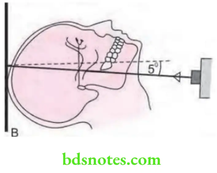
Position of Patient
Head of the patient is centered over the cassette, patient’s head and neck should be tipped back as far as possible, vertex of the skull touches the cassette. Mid-sagittal plane of both the sides is perpendicular to the plane of the film and the radiographic base line is parallel to the film.
Central Ray
Central ray is directed perpendicular to the film and through the mid-sagittal plane, between the angles of the mandible. Central ray should be perpendicular to an imaginary line joining the mandibular fist molars.
“Common challenges in performing the submentovertex view effectively: FAQs provided”
SMV x-ray technique
To view the petrous portion, the central ray should be directed at right angles to the film midway between the external auditory meatus.
Submentovertex View in Radiology: Imaging of the Skull and Mandible
Exposure Parameters
kVp: 50
mA: 20–30
Seconds: 0.4
“Role of proper angulation in capturing the submentovertex view: Questions answered”
Radiographic skull base imaging
Indications
Helps to study destructive/expansile lesions affecting the palate, pterygoid region or base of the skull, sphenoidal sinus.
Leave a Reply