• Skip to main content
  • Skip to secondary menu
  • Skip to primary sidebar
  • About Us
  • Terms of Use
  • Privacy Policy
  • Disclaimer
  • Contact Us
  • Sitemap

BDS Notes

BDS notes, Question and Answers

  • Public Health Dentistry
  • Periodontics
  • Pharmacology
  • Pathology
  • Orthodontics
    • Anchorage In Orthodontics
    • Mandibular Growth, Functional Matrix
    • Retention and Relapse
  • General Surgery
    • Cysts: Types, Causes, Symptoms
    • Maxillofacial Fractures, Disorders, and Treatments
    • Lymphatic Disorders
    • Neurological and Facial Disorders
  • Temporal And Infratemporal Regions
    • Spinal and Neuroanatomy
  • Dental Materials
    • Dental Amalgam
Home » Submandibular Glands

Submandibular Glands

July 25, 2025 by Kristensmith Taylor Leave a Comment

Submandibular Glands

“What are submandibular glands? A detailed question and answers guide”

Question 1. Histology of Submandibular Gland
Answer:

  • Submandibular gland contains serous end peices & mucous tubules
  • Serous end peices contains abundant secretory granules, spherical nucleus & basophilic cytoplasm
  • Mucous secretory cells are filled with pale staining secretory material & little cytoplasm
  • Its nucleus is compressed & contains densely stained chromatin
  • The lumina of mucous tubules are larger
  • The Intercalated & Striated ducts are less in number
  • Connective tissue septa Mucous acini

“Common challenges in mastering submandibular gland notes effectively: FAQs provided”

Blood Vessel Layers Histology

“Understanding submandibular glands through FAQs: Composition, functions, and uses explained”

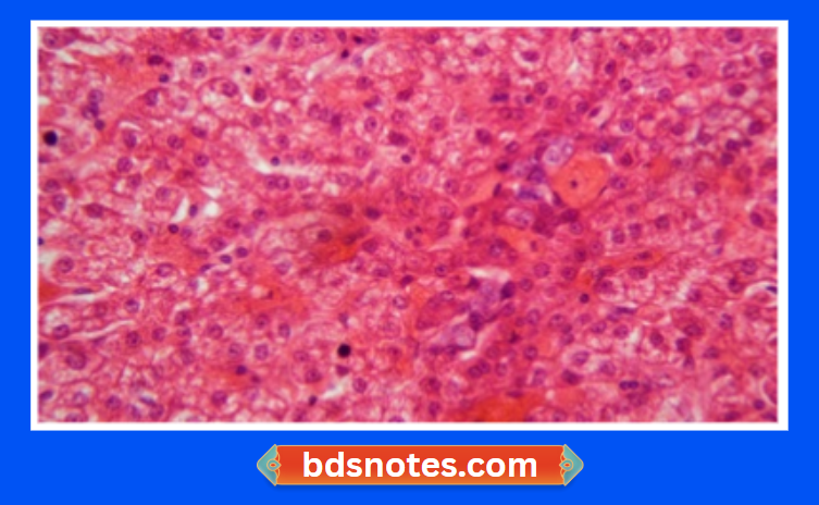
Question 2. Histology of Liver
Answer:

  • The liver is covered by capsule made up of connective tissue
  • The liver substance is divided into large number of lobes consisting of numerous hepatic lobules
  • The lobules appear to merge with one another
  • Each lobule is made up of liver cells called hepatocytes
  • These cells are large with round openfaced nuclei & prominent nucleoli
  • They are separated by sinusoids
  • Sinusoids are surrounded by reticular fibres
  • Along the periphery of each lobules, there are angular intervals called portal canals

“Factors influencing success with submandibular gland studies: Q&A”

    • Each canal contains
    • Branch of portal vein
    • Branch of hepatic artery
    • Interlobular bile duct
  • These structures collectively form portal triad

“Importance of studying submandibular glands for medical students: Questions explained”

Microscopic View Of Liver Tissue

Filed Under: Anatomy

Reader Interactions

Leave a Reply Cancel reply

Your email address will not be published. Required fields are marked *

Primary Sidebar

Recent Posts

  • Branchial Cleft Cyst: Background, Pathophysiology, Etiology
  • Maxillary Nerve: Origin, Course, And Branches
  • The Father Of Anatomy And A Great Anatomist Herophilus
  • Bone Structure – Anatomy
  • The External Carotid Artery: Anatomy, Branches, And Functions
  • Occipitofrontalis Muscle
  • Superficial Temporal Artery
  • Platysma Muscle
  • Cartilage
  • Cauda Equina And Conus Medullaris Syndromes
  • Subcutaneous Injections And Device Management
  • Types Of Circulation: Pulmonary, Systemic, And Portal
  • Structure Of Skeletal Muscle
  • Elastic Cartilage
  • Cellular Organelles And Structure
  • The Golgi Apparatus – The Cell
  • The Cytoplasmic Inclusions Of Certain Plant Cells
  • Dental Abscess
  • Laser Surgery
  • Our Facial Muscles And Their Functions

Copyright © 2026 · Magazine Pro on Genesis Framework · WordPress · Log in