Give a short account of parts, relation & nerve supply of lacrimal gland (or) Nasolacrimal apparatus (or) Name the structures forming lacrimal apparatus
Answer:
Lacrimal Gland Parts
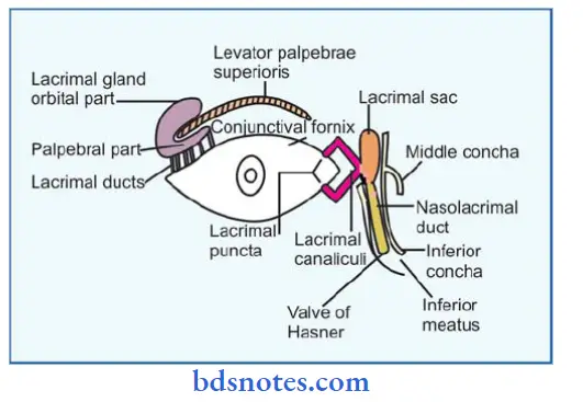
1. Lacrimal gland and its duct:
- Lacrimal gland:
- Lacrimal gland is serous ‘J’ shaped gland
Lacrimal gland Site: - In the lacrintal fossa on the anterolateral part of the roof of the bony orbit & partly on the upper eyelid
Lacrimal gland Parts: - Orbital part: larger & deeper
- Palpebral part: smaller & superficial
- Lacrimal gland is serous ‘J’ shaped gland
- Lacrimal duct:
- Lacrimal duct pierces the conjunctiva of the upper eyelid, open into the conjunctival sac at the superior fornix
- Most of the ducts of orbital part pass through the palpebral part
“Understanding the structures forming the lacrimal apparatus through FAQs: Anatomy, functions, and uses explained”
Conjunctival Sac
- Palpebral conjunctiva:
- Lines the deep surface of eyelids
- Palpebral conjunctiva is thick, opaque, highly vascular & adherent to tarsal plate
- Bulbar conjunctiva:
- Lines the front of eyeball
- Bulbar conjunctiva is thin, transparent & loosely attached to eyeball
- Conjunctival Ssac is the potential space between bulbar & palpebral part
- Conjunctival fornices:
- The lines along which the palpebral conjunctiva of the upper & lower eyelids is reflected on eyeball
“Importance of studying the lacrimal apparatus for medical students: Questions explained”
Lacrimal puncta & canaliculi
- Lacrimal canaliculi is 10 mm long structure beginning at lacrimal punctum
- Lacrimal canaliculi has
- 2 mm long vertical part
- 8 mm long horizontal part
- Lacrimal canaliculi has dilated ampulla at the bend
- Opening
- In the lateral wall of the lacrimal sac behind medial palpebral ligament
4. Lacrimal Sac:
- Lacrimal Sac Site: lacrimal groove behind medial palpebral ligament
- Lacrimal Sac Size: 12 mm long & 5 mm wide
Lacrimal Sac Parts: - Upper end is blind
- Lower end continuous with nasolacrimal duct
Lacrimal Sac Relations: - Anteriorlymedial palpebral ligament, orbicularis oculi
- Mediallylacrimal groove
- Laterallylacrimal fascia & lacrimal part of orbicularis oculi
“Common challenges in mastering lacrimal apparatus notes effectively: FAQs provided”
5. Nasolacrimal Duct:
- Nasolacrimal Duct is 18 mm long membranous passage
Nasolacrimal Duct Course: - Begins at lower end of lacrimal sac
- Runs downwards, backwards & laterally
- Opens into inferior meatus of nose
Valve of Hasner:
- Valve of Hasner is a fold of mucous membrane forming imperfect valve at lower end of duct
Valve of Hasner Nerve Supply:
“Factors influencing success with lacrimal apparatus studies: Q&A”
Leave a Reply