Structure, Distribution, And Clinical Effects of the Ulnar Nerve
Question. Describe the ulnar nerve under the following headings: (a) root value, (b) course and relations, (c) branches and distribution, and (d) applied anatomy.
Answer.
The ulnar nerve is so named because it runs along the ulnar side of the upper limb.
Ulnar nerve
Ulnar Nerve Root value
Ventral rami of C8 and T1. It also gets contribution from the ventral ramus of C7.
Ulnar Nerve Course and Relations
It is the continuation of the medial cord of the brachial plexus in the axilla. It courses successively through four regions: axilla, arm, forearm, and hand, where it terminates by dividing into superficial and deep branches. The course and relations ofthe ulnar nerve in these regions are as follows.
Axilla:
In axilla, the ulnar nerve lies between the axillary vein and the axillary artery on a deeper plane, medial to 3rd part of axillary artery.
Arm
It enters the arm by running downwards on the medial side of the brachial artery in its proximal part. At the midarm (i.e., at the level of insertion of coracobrachialis), it pierces the medial intermuscular septum to enter the back of the arm. Here it descends to run in a groove on the back of the medial epicondyle of the humerus, where it can be palpated.
Forearm:
The ulnar nerve enters the front of the forearm by passing between the two heads of the flexor carpi ulnaris. Here it lies on the medial part of the flexor digitorum profundus. It is accompanied by the ulnar artery on its lateral side in the lower two-thirds of the forearm.
Hand:
The nerve enters the palm by passing superficial to the flexor retinaculum and medial to the ulnar artery. At the distal border of the flexor retinaculum, it ends by dividing into superficial and deep terminal branches.
ulnar nerve damage
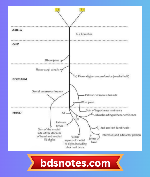
Ulnar Nerve Branches and Distribution
In the axilla and arm:
No branches
In forearm
- Muscular branches to supply:
- Flexor carpi ulnaris
- Flexor digitorum profundus (medial half)
- Palmar cutaneous branch: It arises at about midforearm and provides cutaneous innervation to the skin of the hypothenar eminence.
- Dorsal cutaneous branch: It arises about 5 cm above the wrist and gives off dorsal digital nerves to supply sensory innervation to the dorsal aspects of the medial 1½ digits, excluding their distal phalanges.
In hand
- Superficial terminal branch, which supplies:
- Palmaris brevis muscle
- Cutaneous innervation to the medial one-third of the palm and the medial 1½ fingers, including their nail beds
- Deep terminal branch, which supplies:
- The medial two lumbricals
- Muscles of hypothenar eminence (abductor digiti minimi, flexor digiti minimi, and opponens digiti minimi)
- All the interossei (three palmar and four dorsal)
- Adductor pollicis
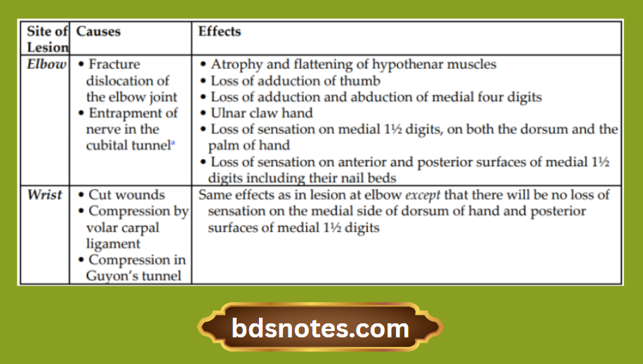
Ulnar Nerve Applied Anatomy
The effects of the lesion on the ulnar nerve depend on the site of the lesion.
Effects of the Ulnar Nerve Lesions
Leave a Reply