• Skip to main content
  • Skip to secondary menu
  • Skip to primary sidebar
  • About Us
  • Terms of Use
  • Privacy Policy
  • Disclaimer
  • Contact Us
  • Sitemap

BDS Notes

BDS notes, Question and Answers

  • Public Health Dentistry
  • Periodontics
  • Pharmacology
  • Pathology
  • Orthodontics
    • Anchorage In Orthodontics
    • Mandibular Growth, Functional Matrix
    • Retention and Relapse
  • General Surgery
    • Cysts: Types, Causes, Symptoms
    • Maxillofacial Fractures, Disorders, and Treatments
    • Lymphatic Disorders
    • Neurological and Facial Disorders
  • Temporal And Infratemporal Regions
    • Spinal and Neuroanatomy
  • Dental Materials
    • Dental Amalgam
Home » Structure And Function Of The Eyes – Eye Disorders

Structure And Function Of The Eyes – Eye Disorders

February 5, 2025 by Kristensmith Taylor Leave a Comment

Structure And Function Of The Eyes – Eye Disorders

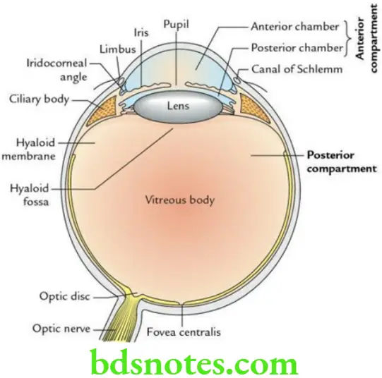
Compartments Of Eyeballs

The interior of the eyeball is divided into two compartments by the lens within the eyeball.

“How does the eye refract light and form images? FAQ answered”

Head And Neck Ear and orbit Compartments of the eyeball

“Understanding the anatomy and function of the eyes through FAQs: Q&A explained”

Anterior compartment: It is small and lies in front of the lens. It is filled with aqueous humour. It is further subdivided into two parts:

  • A smaller anterior chamber, between the iris and cornea.
  • A larger posterior chamber, between the iris and lens.

These two portions of the anterior chamber communicate with each other through a circular aperture in the iris, the pupil.

“Importance of studying the structure and function of the eyes for medical students: Questions explained”

Posterior compartment: It is large (four-fifths) and lies behind the lens. It is filled with a colourless, transparent jelly-like material called vitreous humour/vitreous body.

  • The vitreous humour is enclosed in a delicate hyaloid membrane.
  • Anteriorly, the vitreous body presents a shallow depression (hyaloid fossa) to accommodate the lens.

Filed Under: Head And Neck

Reader Interactions

Leave a Reply Cancel reply

Your email address will not be published. Required fields are marked *

Primary Sidebar

Recent Posts

  • Branchial Cleft Cyst: Background, Pathophysiology, Etiology
  • Maxillary Nerve: Origin, Course, And Branches
  • The Father Of Anatomy And A Great Anatomist Herophilus
  • Bone Structure – Anatomy
  • The External Carotid Artery: Anatomy, Branches, And Functions
  • Occipitofrontalis Muscle
  • Superficial Temporal Artery
  • Platysma Muscle
  • Cartilage
  • Cauda Equina And Conus Medullaris Syndromes
  • Subcutaneous Injections And Device Management
  • Types Of Circulation: Pulmonary, Systemic, And Portal
  • Structure Of Skeletal Muscle
  • Elastic Cartilage
  • Cellular Organelles And Structure
  • The Golgi Apparatus – The Cell
  • The Cytoplasmic Inclusions Of Certain Plant Cells
  • Dental Abscess
  • Laser Surgery
  • Our Facial Muscles And Their Functions

Copyright © 2026 · Magazine Pro on Genesis Framework · WordPress · Log in