Squamous Cell Carcinoma of the Oral Cavity: Classification, Staging, and Grading
Question. Enumerate the carcinomas of the oral cavity. Describe the staging and grading of squamous cell carcinoma.
Answer.
“Importance of studying classification, staging, and grading for better outcomes: Questions explained”
Enumeration of carcinomas of the oral cavity
- Squamous cell carcinoma
- Verrucous carcinoma
- Basaloid squamous cell carcinoma
- Adenoid squamous cell carcinoma
- Spindle cell carcinoma
- Adenosquamous carcinoma
- Undifferentiated carcinoma.
“Understanding squamous cell carcinoma through FAQs: Classification, staging, and grading explained”
Staging of squamous Cell Carcinoma
Staging is defined as extent of spread of tumor within the body.
Staging of squamous cell carcinoma is done by TNM
classification which was given by American Joint Committee
on Cancer (AJCC)
T is suggestive of primary tumor
N is suggestive of regional lymph nodes
M is suggestive of distant metastasis
“Common challenges in diagnosing squamous cell carcinoma effectively: FAQs provided”
T primary tumor
TX Primary tumor cannot be assessed.
T0 No evidence of primary tumor
Tis carcinoma in situ
T1 Tumor 2 cm of less in greatest dimension
T2 Tumor more than 2 cm but not more than 4 cm in greatest dimension
T3 Tumor more than 4 cm in greatest dimension
T4a (Lip) Tumor invades through cortical bone, inferior alveolar nerve, flor of mouth or skin (chin or nose).
T4a (Oral Cavity) Tumor invades through cortical bone, intodeep/extrinsic muscle of tongue (genioglossus, hyoglossus,palatoglossus and styloglossus), maxillary sinus or skin of face.
T4b (lip and oral cavity) Tumor invades masticatory space,pterygoid plates or skull base or encases internal carotid artery
N Regional lymph nodes
NX Regional lymph nodes cannot be assessed
N0 No regional lymph node metastasis
N1 Metastasis in a single ipsilateral lymph node, 3 cm or less in greatest dimension.
N2a Metastasis in a single ipsilateral lymph node, more than3 cm but not more than 6 cm in greatest dimension.
N2b Metastasis in multiple ipsilateral lymph nodes, not more than 6 cm in greatest dimension.
N2c Metastasis in bilateral or contralateral lymph nodes, not more than 6 cm in greatest dimension.
N3 Metastasis in a lymph node more than 6 cm in greatest dimension.
“Factors influencing success with squamous cell carcinoma treatment: Q&A”
M Distant Metastasis
MX Distant metastasis cannot be assessed
M0 No distant metastasis
M1 Distant metastasis.
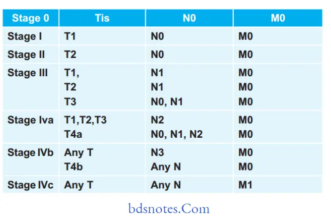
Stage Grouping Of Oral Cancer
“Steps to explain causes of oral squamous cell carcinoma: Tobacco vs alcohol use: Q&A guide”
Grading Of Squamous Cell Carcinoma
- Grading is defined as macroscopic and microscopic degree of differentiation of a tumor.
- Squamous cell carcinoma is divided in following categories by Broder also known as Broder’s classification:
“Role of HPV infection in causing oral squamous cell carcinoma: Questions answered”
Broader’s Classification
- Grade I Well differentiated, – <25% undiffrentiated cells
- Grade II Moderately differentiated – <50% undifferentiated cells
- Grade III Poorly differentiated – <75% undifferentiated cells
- Grade IV Anaplastic/Pleomorphic – >75% undifferentiated cells
Leave a Reply