Specimens Tuberculous (Tb) Lymphadenitis
1. What is this specimen?
- Specimen of lymph nodes that are matted. The cut surface shows caseation. Hence, it is tuberculous lymphadenitis.

“What is tuberculous lymphadenitis?”
2. What is the microscopic picture?
- Central caseation is surrounded by epithelioid cells, Langhans type of giant cells.
3. What are the stages of TB lymphadenitis?
- Stage of lymphadenitis
- Stage of matting
- Stage of cold abscess
- Stage of collar stud abscess
- Stage of sinus formation
4. Why is matting seen in TB lymphadenitis?
- It is because of periodontitis.
5. What is the treatment of cold abscess?
- Nondependent aspiration by using a wide bore needle, to avoid sinus formation.
“Understanding TB lymphadenitis: Causes and symptoms”
Lymphoma
1. What is the diagnosis?
- Multiple lymph nodes which are discrete and not matted. The Cut surface does not show caseation. It is homogenous. Hence, this is a specimen of Hodgkin’s lymphoma.
2. How do you confirm the diagnosis?
- Lymph node biopsy.
3. What is the microscopic picture?
- Cellular pleomorphism: Lymphocytes, histiocytes, eosinophils, monocytes with giant cells containing mirror image nuclei—Reed-Sternberg cell.
4. What are the common lymph nodes involved in Hodgkin’s lymphoma?
- Cervical, axillary, para-aortic, iliac, and inguinal lymph nodes,
5. Is Waldeyer’s ring involvement seen in Hodgkin’s lymphoma?
- No. It is usually seen in non-Hodgkin’s lymphoma.

“Importance of specimen collection in TB lymphadenitis”
Marjolin’s Ulcer
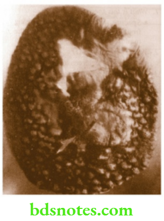
1. What is this specimen?
- Wide excision specimen, showing ulcerated growth arising from the scar. It has everted edges, and there is extensive scarring.
2. What is the diagnosis?
- Squamous cell carcinoma arising in scar tissue is called Marjolin’s ulcer
3. What are the common causes of Marjolin’s ulcer?
- Burns, snake bite, and varicose ulcer
“Complications of untreated TB lymphadenitis”
4. What are the peculiarities of Marjolin’s ulcer?
- It grows very slowly because of scar tissue.
- It is painless as nerves have been destroyed.
- It does not spread by lymphatics as they are also destroyed.
5. What is the treatment?
- Wide excision followed by split skin grafting.
“Common types of specimens used for TB lymphadenitis diagnosis”
Squamous Cell Carcinoma
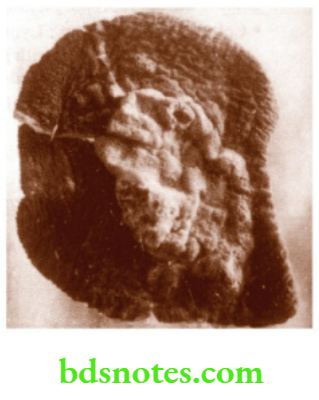
1. What is this specimen?
- Specimen of wide excision showing ulcerated growth with everted edges arising from skin.
2. What is the diagnosis?
- Squamous cell carcinoma
3. What is the microscopic picture?
- Mitotic figures with keratin pearls or epithelial pearls.
4. What is the other treatment for squamous cell carcinoma?
- Radiotherapy
5. What are the common causes of squamous cell carcinoma?
- Leukoplakia
- Radiation dermatitis
- Bowen’s disease
- Congenital skin conditions like xeroderma pigmentosa and albinism
- Chronic scar
“Impact of culture tests on TB lymphadenitis diagnosis”
Specimen Of Hemiglossectomy With Hemimandibulectomy
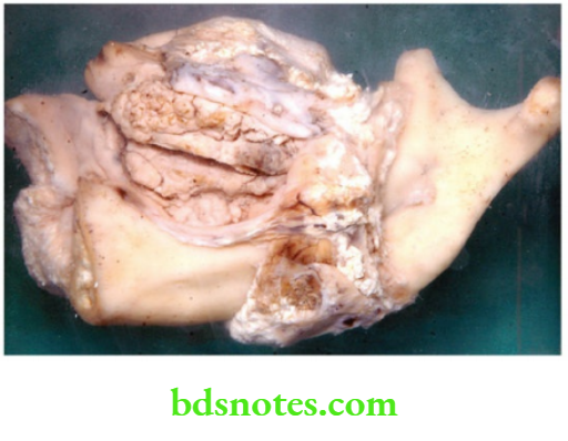
1. What is this specimen?
- Specimen showing growth arising from the tongue and infiltrating the mandible
2. What is the diagnosis?
- Advanced carcinoma tongue
3. Is radiotherapy indicated in this situation?
- No, because the chances of radionecrosis of the mandible are high.
“Role of histopathology in analyzing TB lymphadenitis specimens”
4. What type of X-ray is taken to look for involvement of the mandible?
- Orthopantomogram
5. What is Commando’s operation?
- Hemiglossectomy with excision of the floor of the mouth, hemimandibulectomy, with radical block dissection of the neck done in a single stage, with en bloc removal.
Leave a Reply