• Skip to main content
  • Skip to secondary menu
  • Skip to primary sidebar
  • About Us
  • Terms of Use
  • Privacy Policy
  • Disclaimer
  • Contact Us
  • Sitemap

BDS Notes

BDS notes, Question and Answers

  • Public Health Dentistry
  • Periodontics
  • Pharmacology
  • Pathology
  • Orthodontics
    • Anchorage In Orthodontics
    • Mandibular Growth, Functional Matrix
    • Retention and Relapse
  • General Surgery
    • Cysts: Types, Causes, Symptoms
    • Maxillofacial Fractures, Disorders, and Treatments
    • Lymphatic Disorders
    • Neurological and Facial Disorders
  • Temporal And Infratemporal Regions
    • Spinal and Neuroanatomy
  • Dental Materials
    • Dental Amalgam
Home » Soft Palate: Anatomical Diagram, Function, And Injuries

Soft Palate: Anatomical Diagram, Function, And Injuries

February 9, 2026 by Kristensmith Taylor Leave a Comment

Soft Palate: Anatomical Diagram, Function, And Injuries

Describe the soft palate in brief.
Answer.

It is a movable, muscular flap suspended from the posterior border of the hard palate. It separates the nasopharynx from the oropharynx.

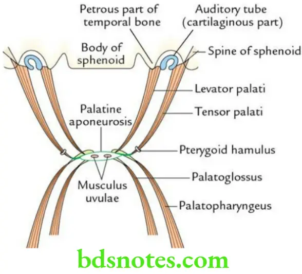
Muscles of the soft palate Palate has five pairs of muscles:

  1. Tensor palati
  2. Levator palate
  3. Musculus uvulae
  4. Palatoglossus
  5. Palatopharyngeus

Head And Neck Pharynx and palate Muscles of the soft palate

Muscles of the soft palate Nerve supply All the muscles of the palate are supplied by the cranial root of the accessory nerve (CN 11) via pharyngeal plexus, except the tensor palate which is supplied by the mandibular nerve (through nerve to medial pterygoid).

Hard Soft Palate

Muscles of soft palate Applied anatomy The paralysis of the soft palate leads to:

  • Nasal regurgitation of food
  • Nasal twang of voice
  • Flattening of the palatal arch on the side of the lesion
  • Deviation of uvula opposite to the side of lesion

Filed Under: Head And Neck

Reader Interactions

Leave a Reply Cancel reply

Your email address will not be published. Required fields are marked *

Primary Sidebar

Recent Posts

  • Branchial Cleft Cyst: Background, Pathophysiology, Etiology
  • Maxillary Nerve: Origin, Course, And Branches
  • The Father Of Anatomy And A Great Anatomist Herophilus
  • Bone Structure – Anatomy
  • The External Carotid Artery: Anatomy, Branches, And Functions
  • Occipitofrontalis Muscle
  • Superficial Temporal Artery
  • Platysma Muscle
  • Cartilage
  • Cauda Equina And Conus Medullaris Syndromes
  • Subcutaneous Injections And Device Management
  • Types Of Circulation: Pulmonary, Systemic, And Portal
  • Hierarchical Organization Of Skeletal Muscle Tissue
  • Elastic Cartilage Histology Short Note For Medical Exams
  • Cellular Organelles And Structure
  • The Golgi Apparatus – The Cell
  • The Cytoplasmic Inclusions Of Certain Plant Cells
  • Dental Abscess
  • Laser Surgery
  • Our Facial Muscles And Their Functions

Copyright © 2026 · Magazine Pro on Genesis Framework · WordPress · Log in