Skeletal Muscle Tissue Explained: Structure, Fibers, And Function
Histology of Skeletal Muscle
Answer:
- The muscle present in relation to bony skeleton is called skeletal muscle
- Histology of skeletal muscle It is made up of
- Muscle fibres
- They are long & cylindrical
- They are arranged in bundles called fasciculi
- Each muscle fibre is covered by a plasma membrane called sarcolemma
- They contain:
- Nucleus
- Elongated nucleus arranged along periphery
- Cytoplasm
- It is called sarcoplasm
- It contains usual cell organelles present near the nucleus
- It also contains myofibrils
- These fibrils are arranged in groups called the fields of Conheim
- Between the myofibrils there is presence of membrane lined tubes called sarcoplasmic reticulum
- Size of fibres:
- Length30 cm
- Diameter 10 to 60 micrometer
- Nucleus
- Muscle fibres
- Skeletal Muscle Connective tissue
- It supports & unites muscle fibres
- The connective tissue surrounding individual muscle fibre is called endomysium
- The connective tissue surrounding individual fasciculi is called perimysium
- Finally the connective tissue surrounding the entire muscle is called the epimysium
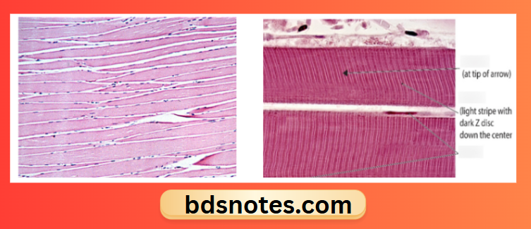
Striations: - The skeletal muscle shows prominent transverse striations
- These striations on staining shows alternate dark & light bands
- A band
- It is dark band
- I band
- Light band of muscle
- Z band
- It is a thin dark line present between I band
- H band
- It is lighter band present between A band
- M Band
- Thin dark line present in center of H band
- The part present between two consecutive Z bands is called Sarcomere.
- A band
Leave a Reply