Sharpey’S Fibers And Tooth Support: Structure, Function, And Clinical Relevance
Question 1. Periodontal ligament.
Answer:
- It is soft, specialized connective tissue situated between the cementum covering the root of the tire tooth and the bone forming the socket wall.
- Width: 0.15 – 0.38 mm.
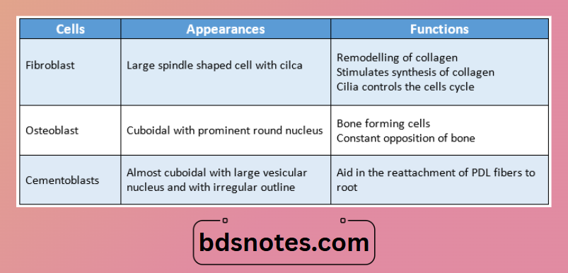
Periodontal ligament Cells:
1. Synthetic cells
- Osteoblasts
- Fibroblast
- Cementoblast.
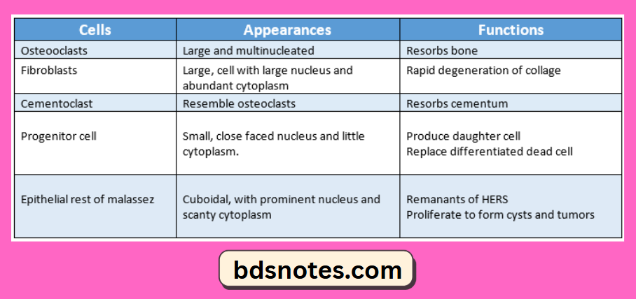
2. Resorptive cells.
- Osteoclast
- Fibroblast
- Cementoclast
3. Progenitor cell.
4. Epithelial rest of malassez.
5. Defense cells.
- Macrophages
- Eosinophils.
Periodontal ligament Functions:
- Supportive
- Sensory
- Nutritive
- Homeostatic
- Eruptive
- Physical
Question 2. Cells of periodontal ligament.
Answer:
Question 3. Sharpey’s fibers.
Answer:
- These are collagen fibers that are embedded into the cementum on one side and into the alveolar bone on another side.
- Fibers in primary acellular cementum are fully mineralized while those in cellular cementum and bone are partly mineralized.
- Their mineralized part appears as a projecting covered with mineral clusters.
- Few of them pass uninterrupted through the alveolar bone to continue as principal fibers of PDL.
- It passes through alveolar bone only when it consists entirely of compact bone.
- It consists of noncollagenous proteins like osteopontin and bone sialoprotein.
Leave a Reply