• Skip to main content
  • Skip to secondary menu
  • Skip to primary sidebar
  • About Us
  • Terms of Use
  • Privacy Policy
  • Disclaimer
  • Contact Us
  • Sitemap

BDS Notes

BDS notes, Question and Answers

  • Public Health Dentistry
  • Periodontics
  • Pharmacology
  • Pathology
  • Orthodontics
    • Anchorage In Orthodontics
    • Mandibular Growth, Functional Matrix
    • Retention and Relapse
  • General Surgery
    • Cysts: Types, Causes, Symptoms
    • Maxillofacial Fractures, Disorders, and Treatments
    • Lymphatic Disorders
    • Neurological and Facial Disorders
  • Temporal And Infratemporal Regions
    • Spinal and Neuroanatomy
  • Dental Materials
    • Dental Amalgam
Home » Salivary Glands – Oral: The Histology Guide – University Of Leeds

Salivary Glands – Oral: The Histology Guide – University Of Leeds

February 9, 2026 by Kristensmith Taylor Leave a Comment

Salivary Glands – Oral: The Histology Guide – University Of Leeds

Describe the histological features of the submandibular salivary gland in brief.
Answer.

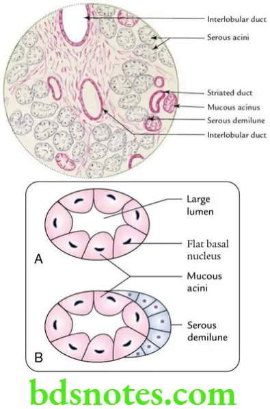
The important histological features of the submandibular salivary gland:

  • Presence of both serous and mucous acini.
  • Mucous acini are made up of truncated columnar cells with flattened basal nuclei. They are stained light pink with H&E.
  • Serous acini are described on p. 120. They stain basophilic in the basal part and pink in the apical part.
  • Serous demilunes of Giannuzzi capping some of the mucous acini are seen.
  • Moderately developed duct system.

Head And Neck Parotid and submandibular regions Histological features of submandibular gland

Filed Under: Head And Neck

Reader Interactions

Leave a Reply Cancel reply

Your email address will not be published. Required fields are marked *

Primary Sidebar

Recent Posts

  • Branchial Cleft Cyst: Background, Pathophysiology, Etiology
  • Maxillary Nerve: Origin, Course, And Branches
  • The Father Of Anatomy And A Great Anatomist Herophilus
  • Bone Structure – Anatomy
  • The External Carotid Artery: Anatomy, Branches, And Functions
  • Occipitofrontalis Muscle
  • Superficial Temporal Artery
  • Platysma Muscle
  • Cartilage
  • Cauda Equina And Conus Medullaris Syndromes
  • Subcutaneous Injections And Device Management
  • Types Of Circulation: Pulmonary, Systemic, And Portal
  • Structure Of Skeletal Muscle
  • Elastic Cartilage
  • Cellular Organelles And Structure
  • The Golgi Apparatus – The Cell
  • The Cytoplasmic Inclusions Of Certain Plant Cells
  • Dental Abscess
  • Laser Surgery
  • Our Facial Muscles And Their Functions

Copyright © 2026 · Magazine Pro on Genesis Framework · WordPress · Log in