Regional Odontodysplasia
Question. Describe histologic features with diagram of ghost teeth.
Answer. It is also known as regional odontodysplasia.
Following are the histologic features of ghost teeth:
“Importance of studying regional odontodysplasia for better diagnostic outcomes: Questions explained”
- In ground section enamel thickness varies.
- Prism structure of enamel is irregular and it lacks laminated appearance.
- Dentin show clefts which are scattred through mixture of interglobular dentin and amorphous material.
- Reduction in amount of dentin is seen.
- Widening of predentin layer is present.
- Large areas of interglobular dentin are seen.
- Pulp contains free or attched pulp stones which exhibit tubules or have laminated calcifiation.
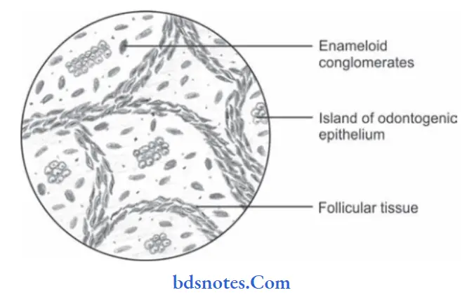
- Follicular tissue surrounding the crown is enlarged and exhibit collections of basophilic enamel like calcifiations called as enameloid conglomerates.
“Understanding regional odontodysplasia through FAQs: Q&A explained”
“Common challenges in diagnosing regional odontodysplasia effectively: FAQs provided”
Leave a Reply