Ramus Of Mandible
Mandible
“What is the ramus of the mandible? A detailed question and answers guide”
Ramus of mandible
Answer:
- It is quadrilateral in shape
- It extends vertically upwards from posterior part of body
Surfaces:
- Lateral
- It is flat & bears a number of oblique ridges
- Medial
- It presents mandibular foramen which leads to mandibular canal
- Anterior margin of it contains tongue shaped projection called lingula
- Mylohyoid groove begins just below mandibular foramen
“Understanding the ramus of the mandible through FAQs: Composition, functions, and uses explained”
Borders:
1. Upper border
- It is thin & curved downwards
- It contains condylar and coronoid process
- Mandibular notch lies between them
- Condylar process
- Its upper expanded part called head joins with fossa of temporal bone to form temporomandibular joint
- Constriction below it is called neck
- Its anterior end presents a depression called the pterygoid fovea
- Coronoid process
- It is flat triangular upward projection
2. Lower border
- It is continuous with base of mandible
- Junction between lower & posterior border forms angle of mandible
“Importance of studying the ramus of the mandible for dental students: Questions explained”
3. Anterior border
- It is thin
4. Posterior border
- It is thick
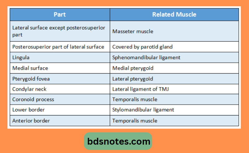
Attachments:
“Common challenges in mastering ramus of the mandible notes effectively: FAQs provided”
Leave a Reply