Radiovisiography
RVG was invented by Dr Frances Mouyens.
- Dr Mouyens invented a way to employ fier optics to narrow down a large X-ray image onto a smaller size that could be sensed by a Charge Coupled Device (CCD) image sensor chip.
- The RVG system is capable of rapidly displaying a digital radiographic image on a monitor which results in a lower patient radiation.
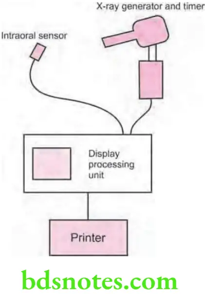
- The “Radio” component is the conventional X-ray generator with a timer, capable of very short exposure time, along with image receptor.
- The “Visio” portion converts the output signal from a CCD to a digital format and displays it on a monitor.
Advantages Of RVG Over Conventional Radiography
- The “Graphy” component consists of data storage unit connected to a video printer.
- The most significant advantages of digital imaging, therefore, are computer-aided image interpretation and image enhancement, in addition to the obvious options of standardized image archiving and image retrieval.
“Early Signs Of Issues With Rvg Imaging”
- The CCD is a solid-state detector composed of an array of X-ray or light sensitive pixels on a pure silicon chip.
- Apixel or picture element consists of a small electron well into which the X-ray or light energy is deposited upon exposure.
- The individual CCD pixel size is approximately 40 μ with the latest versions in the 20μ range. The rows of pixels are arranged in a matrix of 512 x 512 pixels.
- There are two types of digital sensor array designs: area and linear.
- Area arrays are used for intraoral radiography, while linear arrays are used in extraoral imaging. Area arrays are available in sizes comparable to size 0, size 1, and size 2 fims, butthe sensors are rigid and thicker thanradiographic fim and have a smaller sensitive area for image capture.
Clinical Applications Of RVG In Endodontics
- The sensor communicates with the computer through an electrical cable.
“Understanding The Role Of Radiovisiography In Modern Dentistry”
- Area arrays CCDs have two primaryformats: Fiberoptically coupled sensors and direct sensors.
- Fiberoptically coupled sensors utilize a scintillation screen coupled to a CCD. When X-rays interact with the screen material, light photons are generated, detected, and stored by CCD.
- Direct sensor CCD arrays capture the image directly without the intermediate scintillation layer. When exposed to radiation, the covalent bonds between silicon atoms are broken, producing electron whole pairs.
Digital Intraoral Radiography – RVG System
- The number of electron whole pairs that are formed is proportional to the amount of exposure that an area receives. The electrons are then attracted towards the most positive potential in the device, where they create “charge packets”.
- Each packet corresponds to one pixel.
- The charge pattern formed from the individual pixel in the matrix represents the latent image.
“What Is Radiovisiography (Rvg) In Dentistry”
- The image is read by transferring each row of pixel charges from one pixel to the next in a “bucket brigade” fashion. As a charge reaches the end of its row, it is transferred to a read out amplifier and transmitted as a voltage to analog-to-digital convertor located within or connected to the computer.
Benefits Of Radiovisiography In Dental Diagnosis
- Voltages from each pixel are sampled and assigned a numeric value representing a gray level.
“Benefits Of Using Rvg In Dental Diagnostics”
Radiovisiography Clinical applications
- Dental caries detection
- Intrabony defects
- Periapical pathologies detection
- Detection of root fractures
- Detection of root canal length
- Application in mentally retarded/developmentally disabled individuals
- In Telemedicine
Leave a Reply