Radiographic Contrast
Write short note on Radiographic Contrast.
Answer.
Radiographic Contrast
Contrast is the difference in the degree of blackness (densities) between adjacent area on a dental radiography.
- If a dental radiography has very dark areas and very light areas, it is said to have a high contrast, as the dark and the light areas are strikingly different.
- A dental radiograph that does not have very dark and very light areas, but instead has many shades of grey is said to have a ‘low contrast’.
- Radiographic contrast depends on the following factors, i.e.
- Subject contrast
- Film contrast
- Fog and scatter
“Understanding the role of radiographic contrast in imaging: Q&A explained”
Radiographic contrast
Subject Contrast
- Subject contrast refers to the characteristic of the subject which influences the radiographic contrast.
- It depends upon:
- Differences in thickness of subject
- Differences in density of subject
- Differences in tissue atomic number or photoelectric absorption.
“Importance of studying radiographic contrast for better imaging outcomes: Questions explained”
Film contrast
- It is the characteristic of the film which influence the radiographic contrast.
- It is an inherent property of the film itself.
- It determines how the film will respond to the different exposures it receives after the X-ray beam has passed through the patient.
- Film contrast depends upon four factors:
- Characteristic curve of the film
- Optical density or degree of blackening of the film
- Type of film, i.e. direct or indirect action.
- Film processing.
“Common challenges in using radiographic contrast effectively: FAQs provided”
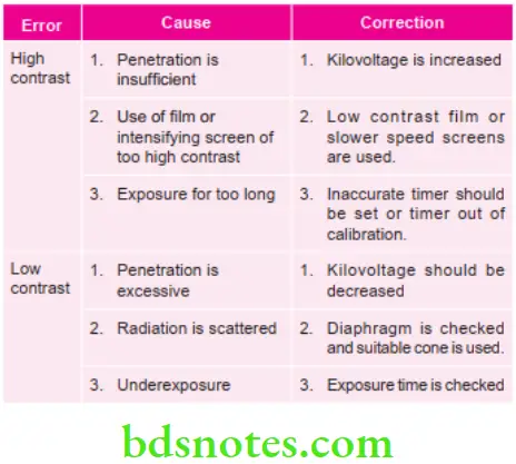
Factors affecting radiographic contrast
Fog and scatter
Radiographic contrast decreases as a result of stray radiation which reaches the film either due to background fog or owing to scatter from within the patient. This produces unwanted film density or darkening.
Radiographic contrast is an exposure and processing error also.
Leave a Reply