Radial Nerve
Question: Describe the radial nerve under the following headings: (a) root value; (b) origin, course, and relations; (c) branches and distribution; and (d) applied anatomy.
Answer.
The radial nerve is the thickest and largest nerve of the upper limb.

Radial Nerve Root value
Ventral rami of C5, C6, C7, C8 and T1.
Radial Nerve Origin, course, and relations
- It arises from the posterior cord of the brachial plexus in the axilla behind the third part of the axillary artery. It is the thickest and largest branch of the brachial plexus.
- It courses successively through three regions: the axilla, the radial groove on the back of the arm, and the front of the forearm. On the front of the forearm, it ends by dividing into superficial and deep terminal branches. The course and relations of the radial nerve in the three regions traversed by it are as follows.
Radial Nerve Axilla:
In the axilla, the radial nerve lies against the muscles forming the posterior wall of the axilla, i.e., subscapularis, teres major, and latissimus dorsi. Then it passes through the lower triangular space between the teres major, the long head of triceps brachii, and the shaft ofthe humerus. In the axilla, it gives two muscular branches to supply the long and medial heads of the triceps and one cutaneous branch (posterior cutaneous nerve of the arm).
radial nerve
Radial groove
The radial nerve from the axilla enters the radial groove through the lower triangular space, where it lies between the long and medial heads of triceps brachii along with the profunda brachii artery. It leaves the radial groove by piercing the lateral intermuscular septum. In the radial groove, it gives three muscular branches to supply the long and medial heads of triceps and anconeus, and two cutaneous branches, i.e. lower lateral cutaneous nerve of the arm and the posterior cutaneous nerve of the forearm.
Radial Nerve Front of arm:
The radial nerve enters the lower anterolateral part of the front of the arm and lies between brachialis on the medial side and brachioradialis and extensor carpi radialis longus on the lateral side. It supplies all these muscles.
Forearm:
The radial nerve enters the cubital fossa where in front of lateral epicondyle it ends by dividing into two terminal branches: (a) superficial terminal branch (superficial radial nerve) and (b) deep terminal branch (posterior interosseous nerve).
Deep terminal branch (posterior interosseous nerve):
It lies in the lateral part of the cubital fossa, where it supplies the extensor carpi radialis brevis and supinator muscles. Then it enters the back of the forearm by passing through the supinator muscle. Here, it supplies abductor pollicis longus, extensor pollicis brevis, extensor pollicis longus, extensor digitorum, extensor indicis, extensor digiti minimi, and extensor carpi ulnaris. At the back of the wrist, it ends in a pseudoganglion, branches of which supply the wrist and distal radioulnar joints.
radial nerve anatomy
Superficial branch (superficial radial nerve):
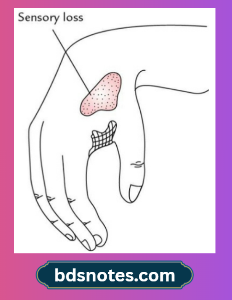
It is regarded as the downward continuation of the trunk of the radial nerve. It runs on the lateral side of the front of the forearm, accompanied by the radial artery in the upper two-thirds of the forearm, with the radial artery being on its medial side. About 7 cm above the wrist, it curves posteriorly deep to the tendon of brachioradialis to reach the anatomical snuff box. Here, it divides into four or five digital branches, which supply the skin of the lateral half of the dorsum of the hand and the lateral 2½ digits till their distal interphalangeal joints.
Radial Nerve Branches and Distribution
In axilla
- Muscular branches: Long and medial heads of triceps brachii
- Cutaneous branches: Posterior cutaneous nerve of the arm
In the radial groove
- Muscular
- Lateral head of triceps brachii
- Medial head of triceps brachii
- Anconeus
- Cutaneous
- Lower lateral cutaneous nerve of the arm
- Posterior cutaneous nerve of the forearm
- Vascular
- To profunda brachii artery
In the arm
- Muscular
- Brachioradialis
- Extensor carpi radialis longus
- Lateral part of brachialis (proprioceptive)
In the forearm
- Superficial terminal branch: Digital branches to supply the skin of the lateral half of the dorsum and the lateral 3½ digits up to the distal interphalangeal (DIP) joints.
- Deep terminal branch (posterior interosseous nerve): Muscular branches to all the muscles of the back of the forearm except anconeus, brachioradialis, and extensor carpi radialis longus.
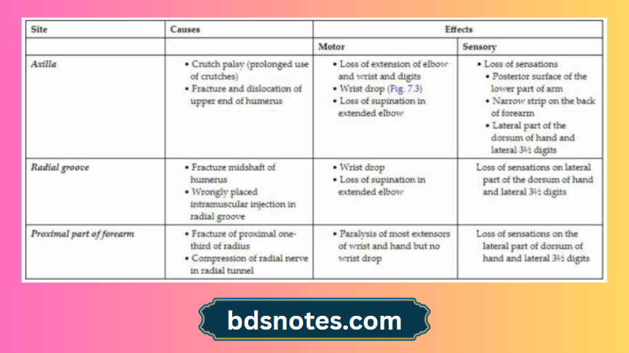
Radial Nerve Applied Anatomy
The effects of injury to the radial nerve at different levels.
Sites of Radial Nerve Injury and Their Effects
Leave a Reply