• Skip to main content
  • Skip to secondary menu
  • Skip to primary sidebar
  • About Us
  • Terms of Use
  • Privacy Policy
  • Disclaimer
  • Contact Us
  • Sitemap

BDS Notes

BDS notes, Question and Answers

  • Public Health Dentistry
  • Periodontics
  • Pharmacology
  • Pathology
  • Orthodontics
    • Anchorage In Orthodontics
    • Mandibular Growth, Functional Matrix
    • Retention and Relapse
  • General Surgery
    • Cysts: Types, Causes, Symptoms
    • Maxillofacial Fractures, Disorders, and Treatments
    • Lymphatic Disorders
    • Neurological and Facial Disorders
  • Temporal And Infratemporal Regions
    • Spinal and Neuroanatomy
  • Dental Materials
    • Dental Amalgam
Home » Pyramidal Tract Explained: From Brain To Spinal Cord

Pyramidal Tract Explained: From Brain To Spinal Cord

February 6, 2026 by Kristensmith Taylor Leave a Comment

Pyramidal Tract Explained: From Brain To Spinal Cord

Describe pyramidal tract with the help of diagram from origin to termination. (or) Draw and label pyramidal pathway.
Answer:

Pyramidal tract:

  • These are descending tracts concerned with voluntary activities of the body.
  • They are also called corticospinal tracts.
  • They are two such tracts.
    • Anterior corticospinal tract.
    • Lateral corticospinal tract.
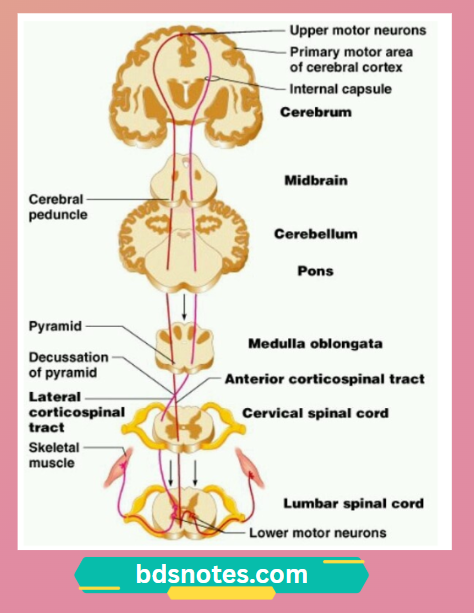
  • The fibres of this tract arises from pyramid like structure in the medulla, thus they are called pyramidal tracts.

Origin:

  • Fibers of pyramidal tract arises from following.

Pyramidal Tract Brain Areas

Course:

  • The fibers of the pyramidal tract descend downwards, in diffused manner and then converge in the form of a fan like structure towards the brain stem as a radiating mass of fibers.
  • This radiating mass is called corona radiata.
  • Then, these fibers reach thalamus and descends down through.

1. Internal capsule:

  • Here, it lie in the genu and anterior two-third of the posterior limb.
  • Then, it enters mid-brain.

2. In midbrain:

  • Here, it lie ventral to the substantia nigra.

3. In pons:

  • Here, the fibers are broken up into a series.
  • At the lower border of pons, the fibers are grouped once again.

4. In the medulla:

  • The fibers occupies the most ventral part of the medulla.
  • Here, the bundle of fibers gives an appearance of the pyramid.
  • At the lower border of medulla, the pyramidal tract on each side is divided into 2 bundles of unequal sizes.

80% of fibers:

  • Cross over and descend as the lateral corticospinal tract and end on the anterior horn cells.
  • While crossing the midline, the fibers form the pyramidal decussation.

20% of fibers:

  • Remain uncrossed
  • Runs downwards in spinal cord as anterior corticospinal tract.

Termination:

  • The fibers from both the tract terminate in the motor neurons of anterior grey horn.
  • The axon of the anterior motor neurons leave the spinal cord as spinal nerves and supply the skeletal muscles.

Corticospinal Tract Pathway

Filed Under: Anatomy

Reader Interactions

Leave a Reply Cancel reply

Your email address will not be published. Required fields are marked *

Primary Sidebar

Recent Posts

  • Branchial Cleft Cyst: Background, Pathophysiology, Etiology
  • Maxillary Nerve: Origin, Course, And Branches
  • The Father Of Anatomy And A Great Anatomist Herophilus
  • Bone Structure – Anatomy
  • The External Carotid Artery: Anatomy, Branches, And Functions
  • Occipitofrontalis Muscle
  • Superficial Temporal Artery
  • Platysma Muscle
  • Cartilage
  • Cauda Equina And Conus Medullaris Syndromes
  • Subcutaneous Injections And Device Management
  • Types Of Circulation: Pulmonary, Systemic, And Portal
  • Structure Of Skeletal Muscle
  • Elastic Cartilage
  • Cellular Organelles And Structure
  • The Golgi Apparatus – The Cell
  • The Cytoplasmic Inclusions Of Certain Plant Cells
  • Dental Abscess
  • Laser Surgery
  • Our Facial Muscles And Their Functions

Copyright © 2026 · Magazine Pro on Genesis Framework · WordPress · Log in