Prevalence And Characteristic Of Oral Mucosa Lesions
Classify oral mucosal lesions. Describe the clinical features and histopathological features of submucosal fibrosis.
Answer. Classification of oral mucosal lesions
Genodermatous
White sponge nevus
Hereditary benign intraepithelial dyskeratosis
Pachyonychia congenita
Porokeratosis
Darier’s disease
Pseudoxanthoma elasticum.
Non-infective Disease
1. Vesicular:
- Erythema multiforme
- Pemphigus
- Bullous lichen planus.
2. Non-vesicular:
Lichen planus.
3. Collagen disorders:
- Lupus erythematous
- Scleroderma.
4. Degenerative
- Oral submucous firosis.
5. Pigmentation:
- Anemia
- Addison’s disease
- Racial pigmentation
OSMF is defined as “An insidious chronic disease affecting any part of oral cavity and sometime pharynx.
Although occasionally preceded by and/or associated with vesicle formation, it is always associated with juxta-epithelial inflammatory reaction followed by firoelastic changes in lamina propria, with epithelial atrophy leading to stiffness of oral mucosa and causing trismus and inability to eat.” —Pindborg (1966)
submucosal fibrosis Clinical Features
- It is caused during 20 to 40 years of age.
- Females are affcted more than males.
- In OSMF firotic changes are frequently seen in buccal mucosa, retromolar area, vulva, tongue, etc.
- Initially patient complains of burning sensation in the mouth, particularly during taking hot and spicy foods.
- There can be excessive salivation, decreased salivation and defective gustatory sensation.
- In initial phase of disease palpation of mucosa elicits a“wet leathery” feeling.
- In advanced stage, the oral mucosa losses its resilience and become blanched and stif and thereby causing trismus.
- Palpation of mucosa often reveals vertical firous bands.
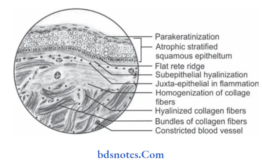
submucosal fibrosis Histopathology
Microscopically OSMF reveals following features:
- Overlying hyperkeratinized, atrophic epithelium often shows flttning and shortening of rete pegs.
- There can be variable degree of cellular atypia or epithelial dysplasia.
- In OSMF dysplastic changes are found in epithelium which include nuclear pleomorphism, severe intercellular edema, etc.
- Stromal blood vessels are dilated and congested and there can be areas of hemorrhage.
- Underlying connective tissue stroma in advanced stage of disease shows homogenization and hyalinization of collagen fiers.
- Decreased number of firoblastic cells and narrowing of blood vessels due to perivascular firosis are present.
- There can be presence of signet cells in some cases.
Leave a Reply