Peripheral Giant Cell Granuloma: Clinical Features, Histology, and Treatment
Question. Write a short note on peripheral giant cell granuloma.
Or
Describe histologic features with a diagram of the peripheral giant cell granuloma.
Answer. Peripheral giant cell granuloma is the most common of giant cell lesions, which arises from tooth-bearing areas of jaw and appears as a purplish red nodule.
“Steps to explain causes of peripheral giant cell granuloma: Trauma vs chronic irritation: Q&A guide”
Peripheral Giant Cell Granuloma Clinical Features
- Lesion usually arises during mixed dentition or during third and fourth decade of life.
- It is most common in males.
- Its site in dentulous patient is interdental papilla. Mandible is more frequently affcted than maxilla.
- Peripheral giant cell granuloma appears as a small, exophytic,well circumscribed, pedunculated lesion on gingival surface.
- Color of lesion varies from purplish red to darkish red.
There can be bleeding from the surface either spontaneously or on provocation from instrument. - Sometimes the peripheral cell granuloma can be aggressive in nature and such lesion may attin very large size and involves some teeth.
In some cases the lesion may develop with an ‘hourglass shape.’
PGCG oral cavity
“Understanding peripheral giant cell granuloma through FAQs: Q&A explained”
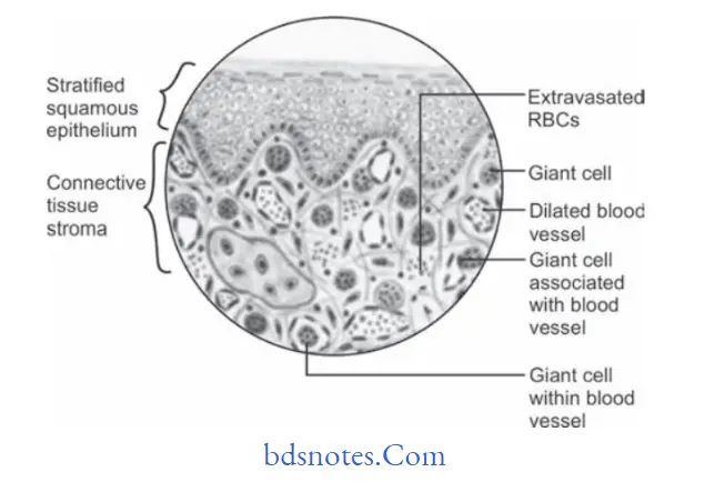
Peripheral Giant Cell Granuloma Histopathology
- Peripheral giant cell granuloma present following histological features.
- Overlying covering epithelium is ulcerated with areas of hemorrhage.
- Underlying connective tissue stroma reveals numerous proliferating firoblasts, blood capillaries and multinucleated giant cells, which are scattred throughout the lesion.
- Fibroblasts present in hypercellular stroma are spindle shaped and have oval shaped nuclei.
- Giant cells are large in size and contain more number of nuclei as compared to true giant cell tumor.
- Areas of hemorrhage and hemosiderin pigment are present within connective tissue stroma.
“Importance of studying peripheral giant cell granuloma for better diagnostic outcomes: Questions explained”
“Common challenges in diagnosing peripheral giant cell granuloma effectively: FAQs provided”
Peripheral Giant Cell Granuloma Treatment
Surgical excision with curettage.
Leave a Reply