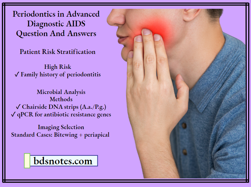
Advanced Diagnostic AIDS
Question 1. Write short note on DNA probe.
Or
Write in detail about advance diagnosis aids in periodontium.
Answer. DNA probes or deoxyribonucleic acid probe:
DNA probes are the advanced diagnostic aids in the microbiological diagnosis.
Probe Design
- DNA probes entails segments of single stranded nucleic acid, labeled with an enzyme.
- A radioisotope, that can be locate and bind to their complementary nucleic acid sequence with low cross reactivity to non-target organisms.
Read And Learn More: Periodontics Question And Answers
Types
- Whole genomic DNA probe.
- Closed DNA probe.
- Synthetic oligonucleotide probe.
Whole Genomic DNA
Whole genomic DNA probes are more likely to cross reaction with nontarget microorganisms due to the presence of homologous sequences between different bacterial species.
Oligonucleotide Probe
Oligonucleotide probes display limited or no cross-reactivity with non-target microorganism.
DNA Probe Procedure
- The plaque is first denaturated to obtain single strain bacterial DNA and then incubate on a membrane such as nitrocellulose. These single stands are individually labeled with a radioactive isotope.
- Specific-labeled DNA probe is incubated on the membrane to allow hybidrization, and then washed off to remove any unhybridized strands.
- The plaque sample contains complementary DNA; hybridization of 2 single strains takes place. It is covered with a radiographic plate.
- The radioactive labels create spots on the film, which are read with a densitometer.
- The darkness and size of the spots indicate the concentration of microorganisms present in the given sample.
DNA Probe Advantages
Sensitivity and specificity of probe are not affected by the presence of unrelated bacteria in the mixed culture sample.
DNA Probe Limitations
Probes are able to detect as few as 102-104 bacteria.
DNA Probe Commercial Kit
Omnigene is the commercially available DNA probe kit for assessment of number of subgingival bacteria.
STEPS IN MICROBIAL DIAGNOSIS WITH DNA PROBE
Question 2. Describe in detail various advanced diagnostic aids.
Or
Write in detail about advanced diagnostic aids in periodontium
Answer. Advanced diagnostic aids are used in periodontics to overcome the problems of conventional diagnostic aids.
Following are the various advanced diagnostic techniques used in periodontology
Advances in Clinical Diagnostic Aids
- Periodontal Probes
- Florida probe system: This automated probe system consists of a probe hand piece, digital readout, foot switch, computer interface, and computer. The end of the probe tip is 0.4 mm in diameter. This probe tip reciprocates through a sleeve, and the edge of the sleeve provides a reference by which measurements are made. These measurements are made electronically and transferred automatically to the computer when the foot switch is pressed. Constant probing force is provided by coil springs inside the probe hand piece and digital readout. This probing method combines the advantages of constant probing force with precise electronic measurement and computer storage of data, thus eliminating the potential errors associated with visual reading and the need for an assistant to record the measurements.
- Periotemp: It enables the calculation of temperature differential between the probed pocket and the subgingival temperature. This temperature differential is useful, because it allows consideration of differences in core temperature between individuals. Elevated subgingival site temperature was particularly related to attachment loss in shallow pockets.
- Periodontal Screening and Recording
- It is designed for easier and faster screening and recording of the periodontal status of a patient or a group of population.
- It uses a especially designed probe that has a 0.5 mm ball tip and is color-coded from 3.5 to 5.5 mm.
- Patient’s mouth is divided into six sextants, i.e. maxillary right quadrant, left quadrant and anteriors, mandibular left and right quadrants and anteriors, and at least six points around each tooth is examined.
- The deepest finding is recorded in each sextant according to the following code.
Code 0: In the deepest sulcus of the sextant, the probes colored band remains completely visible, gingival tissue is healthy and does not bleed on gentle probing. No calculus or defective margins are found. These patients require only appropriate preventive care.
Code 1: The colored band remains completely visible in the deepest sulcus of the sextant. No calculus or defective margins are found but some bleeding after gentle probing is found. Treatment for these patients include subgingival plaque removal and appropriate oral hygiene instructions.
Code 2: The probe’s colored band is still completely visible, but there is bleeding on probing. Supragingival or subgingival calculus and/or defective margins are found. Treatment includes plaque and calculus removal, correction of plaque retentive margins of restorations and oral hygiene instructions.
Code 3: The colored band is partially-submerged. This indicates the need for a comprehensive periodontal examination and charting of the affected sextant to determine the necessary treatment plan. lf two or more sextants score code 3, a comprehensive full mouth examination and charting is indicated.
Code 4: The colored band completely disappears in the pocket, indicating a depth greater than 5.5 millimeters. In this case, a comprehensive full mouth periodontal examination, charting and treatment planning are needed.
Code*: When any of the abnormalities are seen, an asterisk (*) is entered, in addition to the code number.
Advances in Radiographic Diagnostic Aids
- Digital Radiography (RVG):
- Digital radiography allows the use of computerized images, which can be stored, manipulated, and corrected for underexposures and overexposures.
- Digital radiography may yield image properties almost equal to conventional radiographs, but through digital storage and processing, diagnostic information can be enhanced.
- Moreover, there is a one-third to half reduction in radiation dose obtained with digital radiographs compared with conventional radiographs.
- Digital intraoral radiography is in a state of rapid development. Sensors as well as computer hardware and software, are continually modified and improved.
- Because of the clear advantage of real or almost-real images that can be improved and the important educational component of online images presented to the patient, it is expected that digital radiography will soon replace conventional radiography in modern daily practice.
- Computer-assisted densitometric image analysis (CADIA)
- In CADIA, a video camera measures the light transmitted through a radiograph, and the signals from the camera are converted into gray-scale images. The camera is interfaced with an image processor and a computer that allow the storage and mathematic manipulation of the images.
- CADIA appears to offer an objective method for following alveolar bone density changes quantitatively over time. Also, compared with digital subtraction analysis.
- CADIA has shown a higher sensitivity and a high degree of reproducibility and accuracy. This technique has also been applied to longitudinal clinical studies.
- Computerized tomography (CT) scan
- Unlike conventional radiography, which is a two dimensional representation of a three-dimensional object. Computed tomography gives an exact picture of the bone levels in coronal, axial and sagittal plane by which all the osseous defects can be visualized accurately.
- Subtraction radiography: In this procedure, two radiographs are taken and the changes are noted depending on the grey levels.
- Digital subtraction radiography
- Digitalization is done before subtraction, i.e. serial radiographs are converted to digital images.
- Digital images are superimposed and are used on a video screen. Light areas indicate bone gain and dark areas indicate bone loss.
- Absorptiometry: A non-radiographic method to analyze the periodontal bone mass changes. It is based on the absorption by bone of a low energy gamma beam, originating from a radioactive source of l25-I. This method has shown to measure bone changes with a high degree of accuracy and precision.
- Photo-densitometric analysis technique: A beam of light is passed onto the radiographic film and the image is shown on an aluminum scale and then it transforms the density readings into millimeter of aluminum equivalents. It is mainly developed to evaluate bone resorption especially in furcation areas. This technique mainly enables the clinician to detect the variations in the bone density that cannot be detected by visual inspection.
- Nuclear medicine bone scan
- It involves the detection of changes in bone metabolism, so it can detect the earliest stage of bone loss.
- A bone seeking radiopharmaceutical diphosphonate compound is injected intravenously and after the waiting period the uptake by the bone is measured by the semiconductor probe radiation detector.
- This technique is of importance because it detects the bone changes before structural alterations occur.
Advances in Microbiological Diagnostic Aids
- Bacterial culture
- Generally, plaque samples are cultivated under an aerobic conditions, and the use of selective and nonselective media, with several biochemical and physical tests allowing the identification of different putative pathogens.
- The main advantage of this method is that the clinician can obtain relative and absolute counts of the cultured species.
- Moreover, it is the only in vitro method able to assess for antibiotic susceptibility of the microbes.
- Direct microscopy
- Specimens are viewed directly under the light. They are of two types:
- Light microscopy: Under this, stained or unstained specimens can be read.
- Gram’s staining: Differentiates Gram-positive and Gram-negative organisms. Gram-positive appears violet.
- Gram-negative appears pink under the micro-scope.
- This may be important because it differentiates between health and disease.
- Dark field and phase contrast microscopy: Fresh, unstained samples are examined. It uses a special condenser in which the light rays are either reflected or refracted off bacterial cell surface. So the outline of the bacterium is dark against the light background in phase contrast microscopy and light against a dark background in dark-field microscopy.
- Advantages of direct microscopy: It is quick, easy and inexpensive means of screening a microbial sample for major morphotypes.
- Disadvantages:
- Inability to identify species.
- Specimens have to be examined as soon as they are collected from the patients.
- Light microscopy: Under this, stained or unstained specimens can be read.
- Specimens are viewed directly under the light. They are of two types:
- Enzymatic methods
- Tannerella forsythia (Tf), Porphyromonas gingivalis (Pg), the small spirochete Treponema denticola (Td), and Capnocytophega species share a common enzymatic profile: all have a trypsin-like enzyme.
- The activity of this enzyme can be measured with the hydrolysis of the colorless substrate N-benzoyl-DLarginine-naphthylamide (BANA).
- When the hydrolysis takes place, it releases the chromophore 5-naphthylamide, which turns orange red when a drop of fast garnet is added to the solution. Diagnostic kits have been developed using this reaction for the identification of this bacteria profile in plaque isolates (Perioscan).
- Immunodiagnostic methods
- ELISA
- In this, bacterial antigens are incubated in a well, on a plastic plate to allow coating by the material. After washing to remove the free antigen, the plates are ready for tests. Samples containing suspected antibodies and controls are then incubated in separate wells to allow antibodies bind the antigen on the surface of the wells. After washing to remove unbound serum components, antisera to the antibody is conjugated to either alkaline phosphatase or horseradish peroxidase then incubated in the wells. A positive reaction is visualized by addition of a chromogen which changes from a colorless to colored solution.
- Latex agglutination:
- It is based on the binding of protein to latex. Latex beads are coated with species specific antibody and when these beads come in contact with the microbial cell surface. Antigens crosslinking occurs and its clumping/agglutination is made visible within 2 to 5 minutes.
- Direct and Indirect immunofluorescent assay:
- This method permits the identification of specific bacteria in bacterial smears.
- ELISA
- Direct immunofluorescence:
- Antiserum to a microorganism is conjugated to fluorescein.
- The conjugate is incubated on a clinical smear containing the microorganisms and then washed off.
- The antigen-antibody reactions take place and organism is visualized by its fluorescent outline, when observed under a fluorescent microscope, if the microorganism is not present, it appears dark with no fluorescence.
- Indirect immunofluorescence: It is a two step procedure. Antiserum to the microorganism is incubated on the clinical smear and washed off, then a conjugate of a fluorescent dye and an antiserum to the first antisera are incubated and then washed off.
- Flow cytometry: This is for rapid identification of oral bacteria. This involves labeling bacterial cells from a patient plaque sample with both species specific antibody and a second fluorescein conjugated antibody. The suspension is then introduced into the flow cytometer, which separates the bacterial cells into an almost single cell suspension by means of a laminar flow through a narrow tube. After incubation, the cells are passed through a focused laser beam. The cells then scatter the light at low and wide angles, and the fluorescent emission can be measured by appropriate detectors.
Advances in Characterizing Host Response Aids
- GCF assessment (Periotron)
- At present, the most common way to assess GCF is using the Periotron. This electronic device measures the change in capacitance across the wetted strip, and this change is converted to a digital readout, which can be correlated to the volume of GCF. Researchers have established that the Periotron 6000 achieves the easiest and quickest measurement and shows high correlation with other clinical gingival indices.
- Evaluation of inflammatory mediators and products
- Cytokines are potent local mediators of inflammation that are produced by a variety of cells. Cytokines that are present in GCF and have been investigated as potential diagnostic markers include tumor necrosis factor alpha (TNF-a), interleukin-1a (IL-1a, interleukin-1b, Interleukin-6 (IL-6), and interleukin-8 (IL-8). lL-6, and TNF-a are cytokines produced by a variety of cells at inflamed sites. They are potent immunoregulatory molecules with a variety of biologic effects, including metalloproteinase stimulation and bone resorption; therefore they seem good candidates for markers of disease progression.
- Host-derived enzymes: Enzymes such as matrix metalloproteinases, alkaline phosphatases, tissue inhibitors of matrix metalloproteinases, aspartase aminotransferase are used as potential diagnostic markers.
- Tissue breakdown products: One of the major features of periodontitis is the destruction of collagen and extracellular matrices. The connective tissues of the periodontium are composed of fibrous elements, including proteins such as collagen and elastin, and nonfibrous components, including a variety of glycoproteins (laminin, fibronectin, proteoglycans) as well as minerals, lipids, water, and tissue-bound growth factors. The extracellular matrix of the periodontium is composed of a diverse number of macromolecules; the predominant one is collagen, and the other components include proteoglycan (versican, decorin, biglycan, syndecan) and noncollagen proteins (elastin, fibronectin, laminin, osteocalcin, osteopontin, bone sialoprotein, osteonectin, tenascin). All these matrix components are theoretically detectable and potentially informative in terms of their clinical diagnostic utility.
Question 3. Write short note on various radiographs in diagnosis of periodontal diseases.
Answer.
Radiographs used in Diagnosis of Periodontal Diseases
Conventional Radiographic Methods
-
- Intraoral periapical radiograph
- Bitewing radiograph
- Orthopantomograph
- Xeroradiography
Advanced Radiographic Techniques
- Digital radiography
- Xeroradiography
- CADIA
- CT scan
- Subtraction radiography and computer-assisted subtraction
- Absorptiometry
- Photodensitometric analysis technique
- Nuclear medicine bone scan
Conventional Radiographic Methods
Intraoral Periapical Radiograph
They are helpful in assessing the periodontal status and for detecting the periapical infection.
Bitewing Radiographs
They show both maxillary and mandibular teeth along with interdental alveolar bone. So in periodontal diagnosis, they provide us vital information regarding presence of local irritating factors such as calculus.
Orthopantomograph
They have the tendency to underestimate minor bone changes. Major disadvantages are magnification, unsharpen and distortion. Clarity of panoramic image is obtained from digital machines is superior to conventional machines.
Xeroradiography
It does not involve wet chemical processing or the use of a dark room. Instead of X-ray film, xeroradiography uses a uniformly charged selenium plate held in a light tight cassette. Exposure to X-ray and adequate processing produces a real image on opaque paper, which is viewed by reflected light. It is less expensive.
Advanced Radiographic Techniques
These techniques are developed to enhance the ability to see small changes overtime in the bone. The advanced radiographic techniques are:
- Digital Radiography (RVG):
- Digital radiography allows the use of computerized images, which can be stored, manipulated, and corrected for underexposures and overexposures.
- Digital radiography may yield image properties almost equal to conventional radiographs, but through digital storage and processing, diagnostic information can be enhanced.
- Moreover, there is a one-third to half reduction in radiation dose obtained with digital radiographs compared with conventional radiographs.
- Digital intraoral radiography is in a state of rapid development. Sensors as well as computer hardware and software, are continually modified and improved.
- Because of the clear advantage of real or almost-real images that can be improved and the important educational component of online images presented to the patient, it is expected that digital radiography will soon replace conventional radiography in modern daily practice.
- Computer-assisted densitometric image analysis (CADIA)
- In CADIA, a video camera measures the light transmitted through a radiograph, and the signals from the camera are converted into gray-scale images. The camera is interfaced with an image processor and a computer that allow the storage and mathematic manipulation of the images.
- CADIA appears to offer an objective method for following alveolar bone density changes quantitatively over time. Also, compared with digital subtraction analysis.
- CADIA has shown a higher sensitivity and a high degree of reproducibility and accuracy. This technique has also been applied to longitudinal clinical studies.
- Computerized tomography (CT) scan
- Unlike conventional radiography, which is a two dimensional representation of a three-dimensional object. Computed tomography gives an exact picture of the bone levels in coronal, axial and sagittal plane by which all the osseous defects can be visualized accurately.
- Subtraction radiography: In this procedure, two radiographs are taken and the changes are noted depending on the grey levels.
- Digital subtraction radiography
- Digitalization is done before subtraction, i.e. serial radiographs are converted to digital images.
- Digital images are superimposed and are used on a video screen. Light areas indicate bone gain and dark areas indicate bone loss.
- Digital subtraction radiography
- Absorptiometry: A non-radiographic method to analyze the periodontal bone mass changes. It is based on the absorption by bone of a low energy gamma beam, originating from a radioactive source of l25-I. This method has shown to measure bone changes with a high degree of accuracy and precision.
- Photo-densitometric analysis technique: A beam of light is passed onto the radiographic film and the image is shown on an aluminum scale and then it transforms the density readings into millimeter of aluminum equivalents. It is mainly developed to evaluate bone resorption especially in furcation areas. This technique mainly enables the clinician to detect the variations in the bone density that cannot be detected by visual inspection.
- Nuclear medicine bone scan
- It involves the detection of changes in bone metabolism, so it can detect the earliest stage of bone loss.
- A bone seeking radiopharmaceutical diphosphonate compound is injected intravenously and after the waiting period the uptake by the bone is measured by the semiconductor probe radiation detector.
- This technique is of importance because it detects the bone changes before structural alterations occur.
Question 4. Write short note on PCR.
Answer. Full form of PCR is polymerase chain reaction.
- Process of PCR was first conceptualized by Kary Mullis in 1985. Mullis produced virtually unlimited copies of a specific DNA sequence in test tube.
- PCR has three phases which are carried in a instrument known as thermocycler at different temperatures
- Denaturation (Strand separation): This phase is carried out at 94 to 96°C for 20 to 30 seconds, double helical arrangement of the sample template DNA is denatured yielding single stranded DNA molecule.
- Annealing (Primer binding): This phase allows annealing of primers to the single-stranded DNA template at lower temperature of 68°C for 20 to 40 seconds.
- Extension (New DNA synthesis): Final phase of cycle occur at 72°C; Taq DNA polymerase enzyme is added to the reaction mixture to make primer extend along the length of DNA strand. At the end of this phase, a complementary strand is form for each DNA strand of sample DNA that was denatured.
A standard amplification consists of 25 to 35 cycles resulting in formation of billion copies of original sample DNA which is then visualized. The existence of specific amplification product is usually suggestive of presence of target microorganism. So this method is useful in quantitatively recognize number of periodontopathic bacteria at periodontal site.
Types of PCR
- Simplex
- Nested
- Multiplex
- Real time PCR
Advantages of PCR
- Specific: Helps to identify specific bacteria.
- Relatively sensitive: Single molecule of DNA in sample can be detected. PCR has significant potential to produce millions to billion of copies of specific product for sequencing, cloning and analysis.
- Rapid: The process may take few hours to a day.
- Variety of samples from different sources can be tested simultaneously.
- It can detect and identify even when no viable organisms present in the sample and also organisms which are different to cultivate.
Disadvantages of PCR
- It cannot discriminate between viable and non-viable bacteria.
- Not useful for sensitivity tests guiding for selection of antibiotics.
- As small samples are used for amplification process. If this small quantity of plaque sample does not contain targeted microorganism, assay will not detect it.
- Subgingival plaque contains enzymes which can alter amplification process.
- Require thermocycler, specific primer and reagents.
- It is expensive
- It requires experienced personnel.
Question 5. Write short note on CBCT.
Answer. Full form of CBCT is Cone-beam computed tomography.
- It is an imaging modality which offers significant advantages for evaluation of implant patients.
- CBCT was introduced to dentistry in the late 1990s and several CBCT units are commercially available for imaging of craniofacial complex.
- The X-ray source and the detector are diametrically positioned and make 1800 to 3600degree rotation around the patient’s head within the gantry.
- X-ray beam is collimated and resultant beam is cone or pyramid shaped. Typically, a single complete rotation, 180 to 500 basis projections of the region of interest, is generated.
- Computer uses these images to generate a digital, three dimensional map of face. As this map is generated, multiplanar reconstructions i.e. axial, coronal, sagittal, or oblique sections of various thicknesses can be reconstructed from the data.
- Most important feature of the various CBCT units is the field of view describing the extent of the imaged volume.
- CBCT units are categorized as large field of view (greater than 15 cm), medium field of view (8 to 15 cm), and limited field of view systems (less than 8 cm).
- Large field of view units image a more extensive anatomic area, deliver a higher radiation exposure to the patient, and produce lower-resolution images. Conversely, limited field of view units image a small area of the face, deliver less radiation and produce a higher-resolution image.
- CBCT scans offer several advantages for evaluation of the implant patient, compared with two-dimensional (2D) imaging.
- True cross sections offer a precise and detailed evaluation of the height and width of the alveolar ridge. The images can be adjusted and printed without magnification, facilitating measurements directly on the prints or films with standard rulers (i.e., not magnified).
- Vertical and horizontal rulers adjacent to each section allow clinician to check for magnification and make direct measurements.
- Digital format allows for image enhancement tools, rapid communication between the radiologist and the surgeon, and generation of multiple copies of the images.
- Various anatomic structures can be visualized and analyzed at all three coordinate axes, so that their superoinferior, anteroposterior, and buccolingual location can be identified with precision.
- CBCT images the entire arch, or even both arches, so several edentulous areas can be visualized with a single examination. The bone and soft-tissue contrast and resolution are appropriate for the diagnostic task.
- CBCT scanning is a valuable imaging modality for three dimensional and cross-sectional evaluation of the implant patient.
- CBCT imaging delivers much less radiation exposure to the patient.
Leave a Reply