Periapical Granuloma
Question. Write a short note on pulp polyp.
Or
Write a short note on chronic hyperplastic pulpitis.
Or
Write a short answer on pulp polyp.
Answer.
Treatment
- Elimination of polypoid tissue, following the extirpation of the pulp.
- After removing hyperplastic pulp tissue, bleeding can be stopped by pressure.
- Extraction of the tooth or root canal treatment.
“Understanding periapical granulomas through FAQs: Q&A explained”
Question.5. Describe etiology, histopathology, and clinical feature of periapical granuloma.
Answer. It is also called as chronic apical periodontitis.
Periapical granuloma is a localized mass of granulation tissue around the root apex of nonvital tooth which develop in relation to infection and inflammation.
Etiology
- Extension of pulpal inflammation
- Occlusal trauma
- Orthodontic tooth movements with excessive, uncontrolled force
- Acute trauma due to blows on tooth.
- Spread of periodontal infection in the root apex.
- Perforation of root apex into endodontic therapy.
“Importance of studying periapical granulomas for better diagnostic outcomes: Questions explained”
Clinical Features
- Tooth involves produce sensitivity to percussion which occurs due to edema, hyperemia and inflammation of apical periodontal ligament.
- Mild pain and discomfort in a tooth during chewing solid foods.
- Involved tooth is slightly elongated from the socket.
- Periapical granuloma may be asymptomatic in many cases.
- Tooth may be vital or partially vital in initial stages of development of lesion but in fully developed periapical granuloma the affected tooth is nonvital.
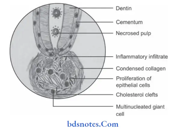
Histopathology
“Common challenges in diagnosing periapical granulomas effectively: FAQs provided”
“Steps to explain causes of periapical granulomas: Pulp necrosis vs bacterial infection: Q&A guide”
- Lesion appears as granulation tissue mass consisting of proliferating fibroblasts, endothelial cells and numerous immature blood capillaries.
- Chronic inflammatory cells, i.e. macrophages, lymphocytes, and plasma cells are present in the lesion.
- There is presence of epithelial islands, cholesterol clefts, and foam cells.
- Plasma cells often produce immunoglobulin there is also present of T lymphocytes in the lesion.
- Epithelial rest cells of Malassez proliferate in response to chronic inflammation, and these proliferating cells undergo certification.
- Bony tissue at the periphery of the lesion is lined by osteoclast cells with an area of bone resorption.
- Few bacteria are present in the lesions which are not affected by the cellular immune mechanism.
- Occasionally, Russell bodies are also found.
- Resorption of cementum and dentin often occurs as a result
of chronic inflammation. In some areas along root, cemento
blastic activity predominates leading to hypercementosis.
Leave a Reply