Parts Of A Dental X-Ray Machine
Draw a neat labeled diagram and explain construction and working of dental X-ray machine.
Answer.
Construction of dental X-ray Machine
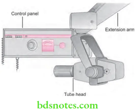
Dental X-ray machine is made up of three parts or components:
- Control panel
- Extension arm
- Tube head
“Understanding the role of each part in a dental X-ray machine: Q&A explained”
Construction of dental X-ray Machine Control panel
It contains
- A on and off switch and an indicator light.
- An exposure button and indicator light.
- Control devices (time, kilovoltage, milliamperage, selectors).
The control panel is plugged into an electrical outlet.
“Importance of studying the parts of a dental X-ray machine for better imaging outcomes: Questions explained”
“Common challenges in identifying parts of a dental X-ray machine: FAQs provided”
Construction of dental X-ray Machine Extension Arm
- It extends from control panel to tube head.
- It houses the electrical wires.
- The extension arm also allows the movement and positioning of tube head.
Dental X-ray machine parts
“Steps to educate patients about dental X-ray machine parts and their importance: Q&A format”
Construction of dental X-ray Machine Tube head
- It is an important part of X-ray machine.
- It contains heavy metal housing, that contains X-ray tube that produce X-rays.
- Following are the components of tube head:
- Metal housing: This is a metal body of tube head that surrounds the X-ray tube and transformer.
- Insulating oil: That surrounds the X-ray tube and transformer inside the tube head, it prevents over heating during X-ray production.
- Tube head seal: Aluminum or leaded glass of the tube head, it act as filter to the X-ray beam and seal the oil in tube head. Permit the exit of X-rays.
- Aluminum disk: Sheets of 0.5 mm thick aluminum is placed in the path of X-ray beam. They act as filter of X-ray beam. In dental X-ray tube head, there are two types of filtration:
Intraoral X-ray machine parts
“Steps to explain the main parts of a dental X-ray machine: Tubehead vs control panel: Q&A guide”
- Inherent filtration: When the primary beam passes through the glass window of the X-ray tube, the insulating oil and the tube head seal. It is approximately equivalent to 0.5 to 1 mm of aluminum.
- Added filtration: Placement of aluminum disks in the path of the X-ray beam between the collimator and the tube head seal in dental X-ray tube head.
- Lead collimator: It is a lead plate with controlled hole. It is used to restrict the size and shape of the X-ray beam. It is placed directly over the opening of the metal housing where X-ray exit. They are of two types, i.e. fixed (dentistry) and adjustable. Lead collimator reduces the patient’s exposure.
- Position indicating device (PID): It is also called “Open ended lead cylinder” or “Cone”. It extends from opening of metal housing of tube head. It appears as an extension of tube head and it aims and shape X-ray beam. They are of following type, i.e. conical, Rectangular and round. Both rectangular and round PID are available in two lengths, i.e. Short (5 inches) Long (16 inches). Long PID is preferred because less divergence of X-ray beam occurs. Rectangular type is most effective in reducing patient’s exposure.
“Differential applications of intraoral vs extraoral X-ray machine parts: Questions answered”
- X-ray tube: It is the main X-ray generating system. It consists of three parts, i.e. lead glass housing, negative cathode and positive anode.
- Lead glass housing is a glass vacuum tube which prevents X-rays in escaping all the directions.
- Negative cathode consists of filament and focusing cup. Filament is basically a coiled wire of tungsten and produces electron on heating. Focusing cup is a holder formed by molybdenum and it houses the filament. Focusing cup focus electrons in a narrow beam and direct beam across the tube towards tungsten target of anode. Cathode supply the electrons which are necessary in generation of X-rays.
- Positive anode converts electrons into X-ray photons.
- Circuits: In X-ray machine two types of circuits are used, i.e. filament circuit or high voltage circuit.
- Filament circuit is of low voltage, i.e. 3 to 5 volts. It regulates flow of electric current to filament of X-ray tube.
- High voltage circuit uses 65,000 to 1,00,000 volts which provide high voltage required to accelerate electrons for generation of X-rays in X-ray tube
- Transformers: It either increases or decrease voltage in an electrical circuit.
- Timer: It completes the circuit with high voltage transformer and controls the time for which high voltage is applied to the tube.
- Tube rating: It is the maximum safe interval the tube is energized at given range of voltage (kVp) and the tube current (mA) values.
- Duty cycle: It refers to how frequently successive exposures can be made.
- X-ray tube: It is the main X-ray generating system. It consists of three parts, i.e. lead glass housing, negative cathode and positive anode.
X-ray machine control panel
“Role of the collimator in a dental X-ray machine: Questions answered”
Working of dental X-ray Machine
- When the X-ray machine is turned on, the electric current enter the control panel; via the plugged in cord and then to the tube head via extension wires in extension arms.
- The current is directed to the filament circuit through the step down transformer, it reduces the 110 to 220 voltage to 3 to 5 volts.
- The filament circuit uses the 3 to 5 volts to heat the tungsten filament. The hot filament emits electron, this emission of electron from the cathode is known as thermionic emission. It forms the electron cloud around the cathode.
- When the exposure button is pushed the high voltage current is activated and the electron cloud is accelerated in X-ray tube from cathode to anode.
X-ray cone dental
“Early warning signs of issues caused by malfunctioning parts of a dental X-ray machine: Common questions”
- Molybdenum cup of cathode directs the electron to the anode target in narrow beam.
- When the electron strikes the tungsten target, their kinetic energy is converted to X-ray energy. Less than 1 % of the energy is converted to X-rays, the remaining 99 % is lost as heat.
- The heat produced is carried away by copper stem and absorbed by the insulating oil in the tube head.
- The area where electron strike on tungsten (anode) is known as focus spot (Tungsten focus).
- Produced X-rays only exit from the X-ray tube via unleaded glass window portion of tube.
- X-rays travel through the unleaded glass window, the tube head seal, the aluminum discs, which filter the long wave X-rays from the beam.
X-ray tube head components
“Asymptomatic vs symptomatic effects of ignoring proper maintenance of X-ray machine parts: Q&A”
- The size and shape of the X-ray beam is controlled by the lead collimator. X-ray beam then travels down the lead lines position indicating device and exit the tube head at the opening of position indicating device.
Leave a Reply