• Skip to main content
  • Skip to secondary menu
  • Skip to primary sidebar
  • About Us
  • Terms of Use
  • Privacy Policy
  • Disclaimer
  • Contact Us
  • Sitemap

BDS Notes

BDS notes, Question and Answers

  • Public Health Dentistry
  • Periodontics
  • Pharmacology
  • Pathology
  • Orthodontics
    • Anchorage In Orthodontics
    • Mandibular Growth, Functional Matrix
    • Retention and Relapse
  • General Surgery
    • Cysts: Types, Causes, Symptoms
    • Maxillofacial Fractures, Disorders, and Treatments
    • Lymphatic Disorders
    • Neurological and Facial Disorders
  • Temporal And Infratemporal Regions
    • Spinal and Neuroanatomy
  • Dental Materials
    • Dental Amalgam
Home » Parotid Gland: Anatomy, Innervation And Clinical Aspects

Parotid Gland: Anatomy, Innervation And Clinical Aspects

February 7, 2025 by Kristensmith Taylor Leave a Comment

Parotid Gland: Anatomy, Innervation And Clinical Aspects

Otic Ganglion Location

“Factors influencing success with parotid gland knowledge: Q&A”

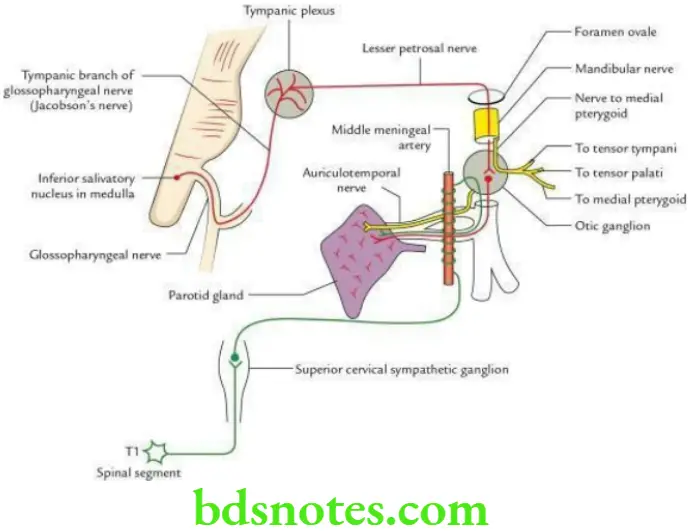
It is a small parasympathetic ganglion of 2–3 mm in size (about the size of pin-head) and is located in the infratemporal fossa, just below the foramen ovale. It lies medial to mandibular nerve and lateral to tensor palati muscle.

Otic Ganglion Roots

Parasympathetic root: From lesser petrosal nerve.

Sympathetic root: From sympathetic plexus around middle meningeal artery.

Sensory root: From auriculotemporal nerve.

Motor root: From nerve to medial pterygoid.

“Understanding the anatomy and innervation of the parotid gland through FAQs: Q&A explained”

Head And Neck Infratemporal fossa temporomandibular joint and pterygopalatine fossa Otic ganglion and its connections

“Importance of studying the parotid gland for medical and dental students: Questions explained”

Otic Ganglion Distribution

Parasympathetic (secretomotor) fibres: Supply parotid gland through auriculotemporal nerve.

Sympathetic (vasoconstrictor) fibres: Supply blood vessels of parotid gland through auriculotemporal nerve.

“Common challenges in understanding parotid gland anatomy effectively: FAQs provided”

Sensory fibres: Provide sensory innervation to parotid gland through auriculotemporal nerve.

Motor fibres: Supply three muscles through nerve to medial pterygoid – medial pterygoid, tensor palati and tensor tympani.

Filed Under: Head And Neck

Reader Interactions

Leave a Reply Cancel reply

Your email address will not be published. Required fields are marked *

Primary Sidebar

Recent Posts

  • Branchial Cleft Cyst: Background, Pathophysiology, Etiology
  • Maxillary Nerve: Origin, Course, And Branches
  • The Father Of Anatomy And A Great Anatomist Herophilus
  • Bone Structure – Anatomy
  • The External Carotid Artery: Anatomy, Branches, And Functions
  • Occipitofrontalis Muscle
  • Superficial Temporal Artery
  • Platysma Muscle
  • Cartilage
  • Cauda Equina And Conus Medullaris Syndromes
  • Subcutaneous Injections And Device Management
  • Types Of Circulation: Pulmonary, Systemic, And Portal
  • Structure Of Skeletal Muscle
  • Elastic Cartilage
  • Cellular Organelles And Structure
  • The Golgi Apparatus – The Cell
  • The Cytoplasmic Inclusions Of Certain Plant Cells
  • Dental Abscess
  • Laser Surgery
  • Our Facial Muscles And Their Functions

Copyright © 2026 · Magazine Pro on Genesis Framework · WordPress · Log in