Parasitology
Question 1. Describe briefly the definitive host
Answer:
Definitive host: When the host harbors the parasite in adult form or when the parasite utilizes the host for sexual reproduction. For example
- In plasmodium: The female anopheles is a definitive host.
- In guinea worm: The man is the definitive host.
Read And Learn More: Microbiology Question And Answers
“Understanding Entamoeba histolytica through FAQs: Q&A explained”
Entamoeba histolytica
Question 2. Describe the life cycle of Entamoeba histolytica and the pathogenesis produced by it.
Answer:
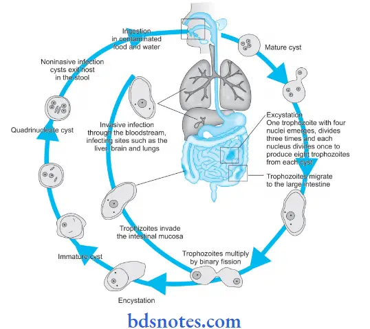
Life Cycle of Entamoeba histolytica:
- E. histolytica passes its life cycle in man.
- Mature quadrinucleate cysts are the infective forms.
- Man acquires the infection by ingestion of water and food containing these cysts.
- Infection can also be acquired by anogenital or orogenital sexual contact among homosexuals.
- As the cyst reaches cecum or lower part of ileum excystation occurs. During this process, each mature cyst liberates a single amoeba with four nuclei, a tetranucleate amoeba that produces eight metacystic trophozoites by division of nuclei by binary fission.
- Liberation of tetranucleate amoeba from cyst occurs due to lysis of the cyst wall by trypsin in the small intestine.
“How does Entamoeba histolytica cause amoebiasis? FAQ answered”
- The meta cystic trophozoites ultimately lodge in the submucous tissue of the large intestine, their normal habitat. Here, they grow and multiply by binary fission.
- During growth, E. histolytica secretes a proteolytic enzyme which brings about the destruction and necrosis of tissues leading to flask-shaped ulcers.
- A large number of trophozoites are excreted along with blood and mucus in the feces. This condition is called amoebic dysentery
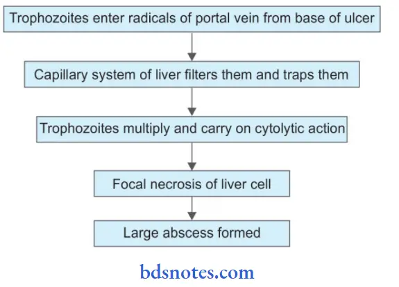
- Sometimes the trophozoites enter into deeper layers and may gain entry into the radicals of the portal vein to be carried away to the liver.
- In the liver, they multiply and produce amoebic hepatitis B and amoebic liver abscesses.
- After some time, when the effect of the parasite on the host is toned down and there is an increase in the tolerance of the host, the lesion starts healing.
- The trophozoites in the lumen of the large intestine, transform into precysts and then into mature quadrinucleate cysts. This process is known as encystation. Cyst formation occurs only within the intestinal tract and not outside the human body.

“Importance of studying parasitology for microbiology students: Questions explained”
Intestinal Lesion of Entamoeba histolytica:
- Acute amoebic dysentery.
- Chronic intestinal amoebiasis.
Patholoenesis of Entamoeba histolytica:

“Common challenges in understanding Entamoeba histolytica effectively: FAQs provided”
Extraintestinal lesions of Entamoeba histolytica:
1. Amoebic liver abscess—pathogenesis

2. Pulmonary amoebiasis—pathogenesis

“Factors influencing success with parasitology knowledge: Q&A”
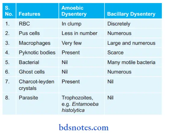
Question 3. Describe briefly the differences between amoebic and bacillary dysentery.
Answer:
Macroscopic:

“Steps to explain the life cycle of Entamoeba histolytica: Cysts vs trophozoites: Q&A guide”
Macroscopic:
 ‘
‘
“Role of cysts in Entamoeba histolytica transmission: Questions answered”
Question 4. Describe the pathogenic lesion produced by Entamoeba histolytica.
Answer:
Pathogenic lesions produced by Entamoeba histolytica
1. Intestinal lesions: Involves large intestine
- Acute amoebic dysentery:
- Complication—multiple ulcers
- Pericecal and pericolic abscess
- Amoebic appendicitis
- Peritonitis
- Perforations
- Gangrene and fitula of gut.
- Chronic intestinal amoebiasis:
- Single latent ulcer in the cecum.
- Multiple small superficial ulcers are scattered throughout the large intestine.
- Thickened cecum and color.
- Pigmented/non-pigmented scar.
2. Extraintestinal or metastatic lesions:
- Liver—amoebic liver abscess
- Multiple small abscesses involving the whole liver.
- A large solitary abscess in the right lobe
- Lung—primary: small multiple abscesses
- Secondary: Single abscess in the lower lobe of the right lung.
- Brain: A small abscess in one of the cerebral hemispheres.
- Spleen: Splenic abscess
- Skin: Granulomatous inflammation of the skin
- Urogenital tract: Amoeba enters through the recto vesicle fistula and rectovaginal fistula.
“Early warning signs of gaps in understanding parasitology basics: Common questions”
Question 5. Write a short note on extraintestinal lesions in amoebiasis.
Answer:
Amoebic Liver Abscess (Hepatic Amoebiasis)
In amoebic liver abscess the:
“Asymptomatic vs symptomatic effects of ignoring parasitology principles: Q&A”
- In this amoeba is transported via the portal circulation to the liver. The invasion of the liver is multifocal and the right lobe is affected commonly.
- Amoebae leads to lytic necrosis and along with an increase in the size of the lesion as well as continuing necrosis there is considerable leukocyte infiltration. Hepatomegaly is present which is known as amoebic hepatitis.
- Lesions can develop in amoebic abscesses. The size of these lesions varies from a few millimeters to several centimeters.
- The Centre of the abscess consists of thick chocolate brown pus with liquefied necrotic liver tissue. Amoeba is located at the periphery.
- Liver abscesses can be solitary or multiple and can cause jaundice.
- If the disease remains untreated some abscesses rupture in adjacent tissue and organ.
Lungs (Pulmonary Amoebiasis):

“Can targeted interventions improve outcomes using Entamoeba histolytica knowledge? FAQs provided”
- This occurs following the rupture of the hepatic abscess via the diaphragm by direct extension.
Brain (Amoebic brain abscess):
- A rare variety of secondary amoebiasis causes amoebic brain abscess which arises as the complication of hepatic or lung abscess or both.
- A small single abscess is located in the cerebral hemisphere.
Spleen (Splenic abscess):
- The involvement of the spleen is due to the liver which causes splenic abscess.
Skin (Cutaneous amoebiasis):
- The skin of the area adjoining the visceral lesion shows sloughing, necrosis, ulceration, and granulomatous mass is known as cutaneous amoebiasis.
“Differential applications of intestinal vs invasive amoebiasis: Questions answered”
Question 6. Write a short note on E. histolytica.
Answer:
E.histolytica is a protozoan: It does not have a field shape and varies due to the extension and retraction of pseudopodia.
Morphology of E. histolytica: It occurs in three stages, which are as follows:
- Trophozoite or Vegetative Form of E. histolytica:
- It has an irregular shape
- Its size is 10 to 40 µm
- This form is actively motile with the help of pseudopodium
- Its cytoplasm is differentiated into ectoplasm which is a thin, clear, translucent outer layer, and endoplasm which is a granular inner layer that consists of a nucleus, food vacuoles, red blood cells, occasionally white cells, and tissue debris.
- Its nucleus is spherical, i.e. of 4 to 6 µ which consists of a small dot-like structure that is central in position and is known as a karyosome. The nucleus is surrounded by a nuclear membrane which is lined with a single layer of uniformly distributed chromatin granules.
- Precystic Stage of E. histolytica:
- This stage occurs during the conversion of a trophozoite to a cyst.
- Its shape is round to oval and the pseudopodium is blunt.
- Its size is 10 to 20 µm
- Its endoplasm is free of red blood cells as well as other ingested particles.
- The nucleus appears the same as seen in the trophozoite stage.
“Steps to master parasitology for exams: Study plans vs mock tests: Q&A guide”
- Cyst Stage of E. histolytica:
- It is spherical in shape.
- Its size is 10 to 15 µm
- Cytoplasm appears as a mass of glycogen and 1 to 4 cigar-shaped or oblong refractile rods known as chromatoidbodies present in immature cysts.
- As the cyst matures the glycogen mass and chromatoid bodies disappear.
- An early cyst consists of a single nucleus. The nucleus undergoes two mitotic divisions to form two and finally four nuclei in mature cysts.
- E. histolytica passes its life cycle in a single host, i.e. man.
- Various methods of reproduction of E. histolytica are encystations and multiplication.
- E. histolytica leads to two types of pathological lesions, i.e. intestinal amoebiasis and extraintestinal amoebiasis.
“Role of diagrams in understanding Entamoeba histolytica mechanisms: Questions answered”
Question 7. Write a short note on extraintestinal amoebiasis.
Or
Write on Extraintestinal Amoebiasis—site and Diagnosis Of Extraintestinal Amoebiasis.
Answer:
Some individuals having intestinal amoebiasis develop hepatic amoebiasis. An amoebic liver abscess is formed. This occurs mostly in the posterolateral surface of the right lobe of the liver.
- The pus of the liver abscess is red-brown in color.
- Pus has closed liver cells, erythrocytes, and leukocytes.
- Pus may liberate trophozoites.
- From the liver, trophozoites may enter into systemic circulation involving other organs such as the lungs, brain, spleen, skin, etc.
Diagnosis Of Extraintestinal Amoebiasis
- Hepatic Amoebiasis:
- Aspiration of pus from liver or liver biopsy should be observed for the trophozoites.
- A stool sample can demonstrate cysts in about 15% of cases.
- Serological tests show the presence of specific antibodies inside the blood.
- Antibodies are detected by various techniques such as complement fixation test, agar gel diffusion, latex agglutination, indirect hemagglutination test, ELISA, immobilization test, and indirect immunofluorescence test.
- Detection of antigen is done by radioimmune assay.
- Amoebic antigens are detected by agglutination tests, ELISA and CIEP.
- Antigen detection is indicative of recent and active infection.
- The polymerase chain reaction is used to detect amoebic DNA in aspirated pus from the amoebic liver abscess.
- Radiographic examination shows raised right dome of the diaphragm.
- Pulmonary Abscess:
- Trophozoites are demonstrated by red-brown anchovy sauce sputum.
“Early warning signs of poor performance in parasitology exams: Common questions”
Question 8. Write short note on amoebiasis.
Answer:
Amoebiasis is a disease caused by the protozoan Entamoeba histolytica Amoebiasis is of two types, i.e. intestinal and extraintestinal.
- Intestinal Amoebiasis:
- It is also known as primary amoebiasis.
- There is a presence of blood and mucus in stool
- The stool is foul-smelling and is brown-black in color.
- The patient is afebrile and is non-toxic.
- At times there is diarrhea and vague abdominal symptoms are present.
- Extraintestinal Amoebiasis:
- Most commonly occurs hepatic amoebiasis which is characterized by:
- Pain as well as tenderness in the right hypochondrium.
- Presence of fever with chills.
- As there is irritation of the phrenic nerve there is the presence of shoulder pain.
- Weight loss is present.
- Infrequent jaundice is present.
- There is also the presence of pulmonary amoebiasis, cutaneous amoebiasis, splenic abscess, and cerebral amoebiasis.
“Can advanced tools supplement parasitology exam preparation? FAQs provided”
Laboratory Diagnosis:
1. Intestinal Amoebiasis:
- Stool examination:
- In acute amoebic dysentery stool or colonic scrapings from ulcers should be examined by both naked eye and microscopic examination.
- Normal saline preparation is used for the demonstration of actively motile trophozoites. Charcot-Leyden crystals can appear in saline preparation.
- Iodine preparation is used to study cysts or dead trophozoites.
- The concentration method, i.e. formal ether can be used for the concentration of amoebic cysts in the stool when amoebae are scanty.
- Stool antigen detection: ELISA is used to detect antigens of E. histolytica in feces.
- Blood examination: This demonstrates leukocytosis.
- Serological tests: Antibodies are visible in the later stages of intestinal amoebiasis.
- Various serological tests used are ELISA, indirect fluorescent antibody, and indirect hemagglutination assay.
- Polymerase chain reaction: In feces, E. histolytica is identified by this technique.
2. Extraintestinal Amoebiasis:
- Hepatic Amoebiasis:
- Aspiration of pus from liver or liver biopsy should be observed for the trophozoites.
- A stool sample can demonstrate cysts in about 15% of cases.
- Serological tests show the presence of specific antibodies inside the blood.
- Antibodies are detected by various techniques such as complement fixation test, agar gel diffusion, latex agglutination, indirect hemagglutination test, ELISA, immobilization test, and indirect immunofluorescence test.
- Detection of antigen is done by radioimmune assay.
- amoebic antigens are detected by agglutination tests, ELISA and CIEP. Antigen detection is indicative of recent and active infection.
- The polymerase chain reaction is used to detect amoebic DNA in aspirated pus from the amoebic liver abscess.
- Radiographic examination shows raised right dome of the diaphragm.
“Asymptomatic vs symptomatic effects of outdated study methods: Answered”
- Pulmonary Abscess:
- Trophozoites are demonstrated by red-brown anchovy sauce sputum.
- Treatment Of Amoebiasis
- Metronidazole should be given in both intestinal as well as extraintestinal amoebiasis.
- Diloxanide furoate, diiodohydroxyquin, and paromomycin is active against trophozoites and cysts.
- In both amoebic colitis and amoebic abscess metronidazole, tinidazole, secnidazole and ornidazole are very effctive.
- In hepatic and pulmonary abscess chloroquine, tinidazole, emetine hydrochloride, tetracycline, etc.
Leave a Reply