• Skip to main content
  • Skip to secondary menu
  • Skip to primary sidebar
  • About Us
  • Terms of Use
  • Privacy Policy
  • Disclaimer
  • Contact Us
  • Sitemap

BDS Notes

BDS notes, Question and Answers

  • Public Health Dentistry
  • Periodontics
  • Pharmacology
  • Pathology
  • Orthodontics
    • Anchorage In Orthodontics
    • Mandibular Growth, Functional Matrix
    • Retention and Relapse
  • General Surgery
    • Cysts: Types, Causes, Symptoms
    • Maxillofacial Fractures, Disorders, and Treatments
    • Lymphatic Disorders
    • Neurological and Facial Disorders
  • Temporal And Infratemporal Regions
    • Spinal and Neuroanatomy
  • Dental Materials
    • Dental Amalgam
Home » Pain Pathways

Pain Pathways

February 6, 2026 by Kristensmith Taylor Leave a Comment

Pain Pathways

Question 1. Describe origin, course and function of corticospinal. (or) Functions of pyramidal tract.
Answer:

  • Pyramidal tract is also called corticospinal tract.

Corticospinal Functions:

1. The pyramidal tracts are concerned with voluntary movement of the body.

  • Lateral corticospinal tract.
    • Convey motor impulses to control voluntary movements specially of the distal limb muscles.
    • Concern with fine, precise movements.
  • Anterior corticospinal tract.
  • Convey motor impulses to control movements of trunk and proximal portions of the limb.

2. Forms part of pathway for superifcial reflexes

3. Fibers of this tract are concerned with sensory motor coordination.

Question 2. Describe the pain pathway. (or) Pathway of pain.
Answer:

  • Lateral spinothalamic tract carries the pain sensation from the peripheral parts of the body to the CNS.

Pain Pathway Origin:

  • Pain fibers arises from free nerve endings that are distributed throughout the body.

Pain Pathway Course:

1. First order neurons – posterior nerve root ganglia cells.

  • Dendrites of these neurons receive the impulses and their axons carry them to the spinal cord.
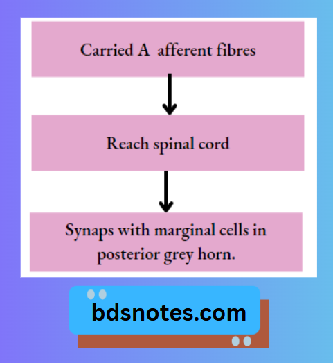
    • Fibers for fast pain.

A Afferent Fibers Spinal Cord

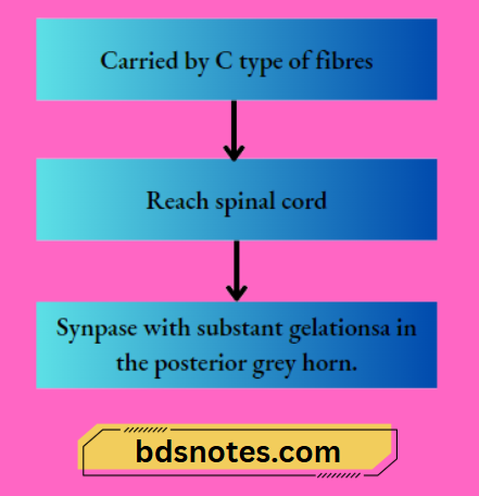
2. Fibers for slow pain.

C Type Fibers Spinal Cord

2. Second order neuron – marginal cells and substantia gelatinosa cells.

1. Fast pain.

Neospinothalamic Tract Formation

2. Slow pain.

Paleospinothalamic Tract Formation

Pain Sensation Pathway Diagram

Pain Pathway Termination:
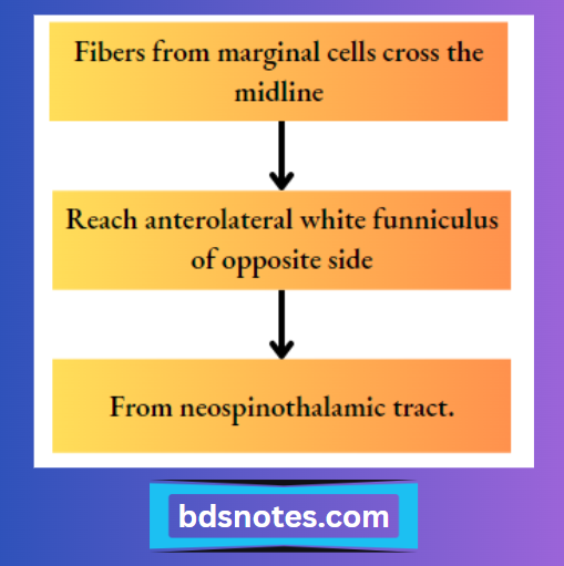
1. Fast pain fibers

  • Majority of fibers terminate in ventral posterolateral nucleus of thalamus.
  • Few fibers terminate in ascending reticular system of brainstem.

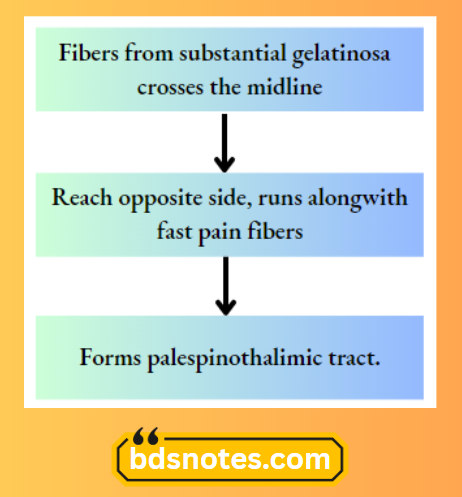
2. Slow pain fibers.

  • One-fifth of fibers terminate in ventral posterolateral nucleus of thalamus.
  • Remaining fibers terminate in either of the following.
    1. Reticular formation of brainstem.
    2. Tectum of midbrain
    3. Grey matter around aqueduct of sylvius.
  • These terminating sites form third order neuron.
  • Axons from these neurons reach the sensory area of the cerebral cortex.

Filed Under: Anatomy

Reader Interactions

Leave a Reply Cancel reply

Your email address will not be published. Required fields are marked *

Primary Sidebar

Recent Posts

  • Branchial Cleft Cyst: Background, Pathophysiology, Etiology
  • Maxillary Nerve: Origin, Course, And Branches
  • The Father Of Anatomy And A Great Anatomist Herophilus
  • Bone Structure – Anatomy
  • The External Carotid Artery: Anatomy, Branches, And Functions
  • Occipitofrontalis Muscle
  • Superficial Temporal Artery
  • Platysma Muscle
  • Cartilage
  • Cauda Equina And Conus Medullaris Syndromes
  • Subcutaneous Injections And Device Management
  • Types Of Circulation: Pulmonary, Systemic, And Portal
  • Structure Of Skeletal Muscle
  • Elastic Cartilage
  • Cellular Organelles And Structure
  • The Golgi Apparatus – The Cell
  • The Cytoplasmic Inclusions Of Certain Plant Cells
  • Dental Abscess
  • Laser Surgery
  • Our Facial Muscles And Their Functions

Copyright © 2026 · Magazine Pro on Genesis Framework · WordPress · Log in