Understanding Osteitis Deformans (Paget’s Disease): From Skull to Jaw
Question: Write a note on Paget’s disease of bone.
Or
Write notes on the histopathology of Paget’s disease.
Or
Write in detail on Paget’s disease.
Or
Write short notea on Paget’s disease.
“Understanding Paget’s disease of the skull and jaw through FAQs: Q&A explained”
Answer. Paaget’s disease is a relatively uncommon bony disorder which is characterized by the excessive uncoordinated phases of resorption and deposition of osseous tissue in single or multiple bones.
It is also known as “Osteitis Deformans.”
Etiology Of Paget’s disease.
- Inflammatory
- Circulatory disturbance
- Genetic and environmental factors
- Others: Vasculitis, trauma, hormonal balance and degenerative neurological disorders.
“Importance of studying Paget’s disease for better diagnostic outcomes: Questions explained”
Clinical Features Of Paget’s disease.
- It occurs during 5th, 6th and 7th decades of life.
- Males are more commonly affcted.
- It is prone to occur in axial skeleton especially in skull,femur, sacrum and pelvis.
- Most of the patient complain initially of the deep and aching bone pain with bilaterally symmetrical swelling of bone.
- Enlargement of skull, bowing of legs and curvature of spine are commonly seen.
Oral Manifestations Of Paget’s disease.
- Maxilla is more commonly involved than the mandible.
- There is movement and migration of affcted teeth and malocclusion.
- As the disease progresses, the mouth remains open exposing the teeth.
- Extraction site heals slowly and incidence of osteomyelitis is higher.
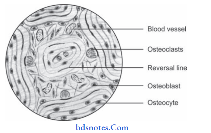
Histopathology Of Paget’s disease.
“Common challenges in diagnosing Paget’s disease effectively: FAQs provided”
- Osteoclastic bone resorption occurs and the bone is replaced by highly vascularized cellular connective tissue.
- Osteoclasts are usually larger and may contain over 100 nuclei.
- Deposition of new lamellar or woven bone within the connective tissue by osteoblast cells occurs and fatty hemopoietic bone marrow is replaced by the fibrous stroma.
- Newly formed bone may again resorbed by osteoclast causing loss of normal architecture of bone.
- Chronic inflammatory cells and dilated blood capillaries are present within the bone.
- Bone resorption and deposition results in irregular fragments of bone formation characterized by prominent basophilic reversal and resting lines, and they produce mosaic pattern in bone.
“Why is early detection critical for managing Paget’s disease? Answered”
“Steps to explain causes of Paget’s disease: Genetic vs environmental factors: Q&A guide”
Treatment Of Paget’s disease.
- Bisphosphonate therapy should be given.
- Calcitonin is administered.
- Surgical correction for bone deformities, fractures and severe degenerative arthritis
Leave a Reply