Oxygen Transport In Blood: From Lungs To Tissues
Describe transport of oxygen in blood. (or) Oxygen-haemoglobin dissociation curve.
Answer:
Transport of oxygen:
- Oxygen is transported by the blood from alveoli to the tissue.
- Oxygen is transported in blood in 2 forms.
1. Dissolved form:
- Oxygen dissolves in water of plasma and is transported.
- Oxygen transported in this form is 0.3 ml per 100 ml of blood per 100 mm Hg pO2
Oxygen in Blood Significance:
- About 3% of total oxygen in blood is transported in this form.
- Oxygen is transported in this form in stress full conditions like exercise.
- It occurs due to excess demand of oxygen by the tissues.
Amount of oxygen transported:
- Arterial – 0.3 ml per 100 ml of blood.
- Venous – 0.12 ml per 100 ml of blood.
2. In combination with haemoglobin:
- Oxygen combines with haemoglobin in blood and is transported as oxyhaemoglobin.
- Each haemoglobin molecule has 4 heme groups which have an iron in ferrous form.
- Sixth valency bond of each Fe2+ combines with 2 atoms of oxygem.
- Therefore, S atoms of oxygen combines with one mole of haemoglobin.
- No oxidation reaction takes place during this combination.
Oxygen in Blood Significance:
- Maximum amount of oxygen, about 97% is transported in this form.
- Oxygen can be released from haemoglobin easily when needed.
- Haemoglobin also accepts oxygen readily when the pO2 is more and gives out oxygen when the pressure is less.
Oxygen carrying capacity of haemoglobin:
- One gram of haemoglobin carried 1.34 ml of oxygen.
- This is called oxygen carrying capacity of haemoglobin.
- 1 gram of Hb = 1.34 ml of oxygen.
- Now, normal haemoglobin content – 15 gram%.
- So, oxygen carried in this form (15 x 1.34) ml = 20.1 ml of oxygen in 100 ml of blood.
- But the 15% of haemoglobin carries only 19 ml% of oxygen.
- It is due to absence of full saturation of haemoglobin with oxygen.
- It is saturated only for about 95%.
- The oxygen carrying capacity of haemoglobin is given by oxygen haemoglobin dissociation curve.
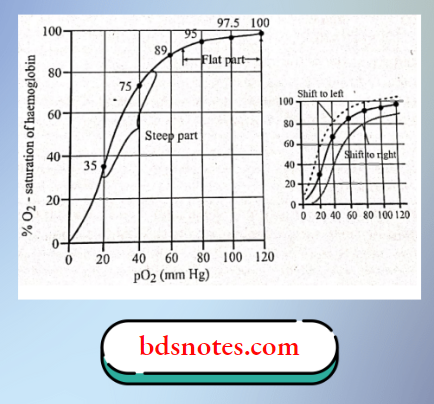
Oxygen haemoglobin dissociation curve:
- The relationship between the partial pressure of oxygen and the percentage saturation of haemoglobin with oxygen is explained graphically by the oxygen haemoglobin dissociation curve.
- It is sigmoid shaped.
- Lower part of curve.
- Indicates dissociation of oxygen from haemoglobin.
- Upper part of curve.
- Indicates acceptance of oxygen by haemoglobin depending upon the partial pressure of oxygen.
- Lower part of curve.
Amount of oxygen transported in this form:
- Arterial blood = 19 ml per 100 ml of blood.
- Venous blood 13.88 ml per 100 ml of blood.
Leave a Reply