Oral Mucous Membrane: Structure, Function, And Specialized Regions
Write about the specialized mucosa of the oral cavity.
Answer:
Specialized mucosa:
- The mucous membrane covering the dorsum of the tongue is called a specialized mucosa.
- It is covered by functionally highly extensible masticatory mucosa.
Dorsum of the tongue:
- It is rough and irregular.
- It is covered by a stratified squamous keratinized epithelium.
- It is divided by the V-shaped groove called sulcus terminals into:
1. Anterior two-thirds- the body of the papillary part.
- Mucosa is derived from the first pharyngeal arch.
- It is supplied by the lingual nerve.
2. Posterior one-third-base or lymphatic part.
- Derived from the third pharyngeal arch.
- It is supplied by the glossopharyngeal nerve.
Fungiform papillae:
- They are scattered between the numerous filiform papillae at the tip of the tongue.
- They are red, smooth, round structures.
- They are covered by thin, nonkeratinized epithelium.
- They have a rich capillary network.
- They contain one to three taste buds only on their dorsal surface.
Filiform Papillae:
- They are fine-pointed, cone-shaped papillae covering the anterior part of the tongue.
- Covered by a thick keratinized epithelium, containing a core of connective tissue.
- They form a tough, abrasive surface.
- They do not contain taste buds.
- They are involved in compressing and breaking food when the tire tongue is opposed to the tire hard palate.
Foliate papillae:
- They are leaflike papillae present on the lateral margins of the posterior part of the tongue.
- They consist of 4-11 parallel ridges separated by deep grooves.
- They contain few taste buds.
- They are more frequent in mammals.
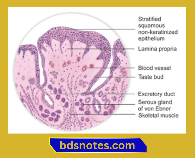
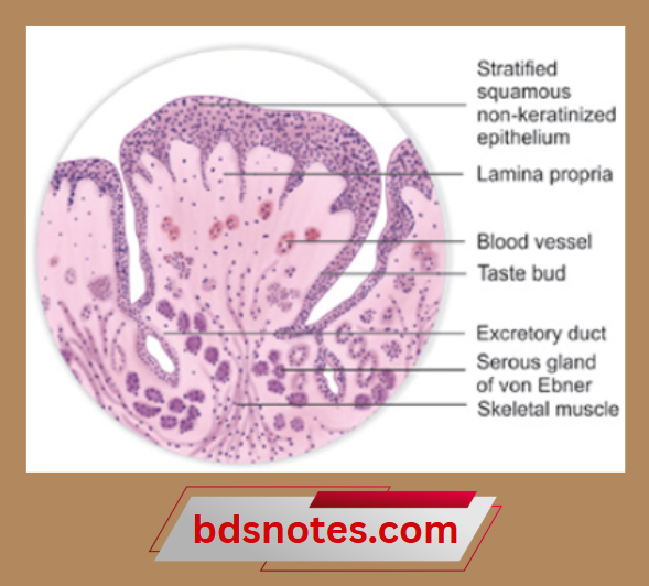
Circumvallate papillae:
- They are 8-12 large structures present just anterior to the sulcus terminalis.
- They are surrounded by a deep, circular groove for the opening of ducts of minor salivary glands.
- They contain a connective tissue core covered by a keratinized epithelium.
- Taste buds are present on its lateral surface.
- Its free surface shows numerous secondary papillae.
- They do not protrude above the surface of the tongue.
- Their function is to wash out the soluble food elements.
- They are also the main source of salivary lipase.
Posterior one-third of the tongue:
- It contains round or oval prominences – lingual follicles.
- Each of these has one or more lymph nodules.
- Lingual follicles have a small central pit called a lingual crypt lined with stratified squamous epithelium.
- Ducts of small mucous lingual glands open into the lingual crypt.
- The lingual follicles together form the lingual tonsil.
Leave a Reply