Oral Cancer
Question.1. Describe the etiology, pathology, clinical features and treatment of oral cancer.
Or
Describe the etiological factors, clinical features and management of oral carcinoma.
Answer.
Etiology
Etiological Factors Of Oral Cancer
Tobacco smoking:
- Cigarettes
- Bidis
- Pipes
- Cigars
- Reverse smoking.
Use of smoking tobacco:
- Snuff dipping
- Tobacco sachets
- Tobacco chewing:
- Betal chewing
- Chewing of areca nut
- Consumption of alcohol
- Diet and nutrition: Vit. A, B complex and C deficiency.
Read And Learn More: General Surgery Question And Answers
Dental factors:
- Chronic irritation from broken teeth.
- Ill-fitting or broken prosthesis.
Ultraviolet radiation: Actinic radiation
Viruses:
- Herpes simplex virus
- Human papilloma virus
- Human immunodeficiency virus (HIV)
- Epstein-Barr virus
Immunosuppression:
- AIDS
- Organ transplantation
Chronic infection:
- Candidiasis
- Syphilis.
Occupational hazards: Woollen textile workers.
Genetic factors:
- Oncogenes
- Tumor suppressor genes
Pre-existing oral disease:
- Oral lichen planus
- Oral sub-mucous firosis
- Leukoplakia.
Oral Cancer Clinical Features
- Male predilection is seen.
- Carcinoma mostly occurs in the older age.
- Site: Most commonly involved are the posterior and lateral borders of tongue and lower lip and less frequently the flor of mouth, palate and buccal mucosa.
- Small lesion is asymptomatic.
- Large lesion may cause some pain or paresthesia and swelling.
- Patients complain of persistent ulcer in the oral cavity.
- Function of organ is impaired.
- Appearance: The clinical appearance of a carcinomatous ulcer is that one of irregular shape induration and raised everted edges.
- Base: Usually have broad base and are dome like or nodular.
- Surface: May range from granular to pebbly to deeply creviced.
- Surface may be entirely necrotic and have ragged whitish gray appearance.
- Color: It may be completely red or red surface may be sprinkled with white necrotic or keratin area.
- Lymph nodes: Superficial and deep cervical nodes are commonly affected.
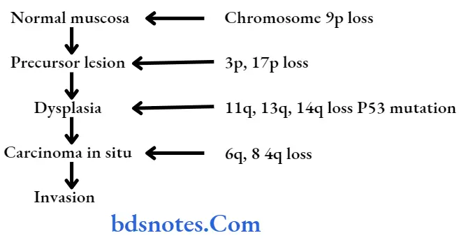
Oral Cancer Pathology
- Allelic imbalance (loss of heterozygosity [LOH]) has been identifid in tumor suppressor gene.
- Damage to tumor suppressor gene may also involve damage to other genes involved in growth control, mainly those involve in cell signaling (oncogenes, especially some on chromosome 11 and chromosome 16)
- Changes in oncogenes can disrupt cell growth control,leading ultimately to uncontrolled growth of cancer.
Management Of Oral Cancer
Management should be curative or palliative:
Early growth without bone involvement
- Curative radiotherapy using caesium needles or iridium wires, i.e. brachytherapy.
- Other option is wide excision wire 1–2 cm clearance.
Often, the approach to the tumor is by raising the cheek flp (outside). After the wide excision, the flap is placed back (Pattrson operation). - Presently advanced technology in radiotherapy facilitates the use of external radiotherapy also.
The incidence of dreaded complication like osteoradionecrosis mandible has been reduced due to bettr radiotherapy methods. - Growth with mandible involvement: Here along with wide excision of the primary tumor hemimandibulectomy or segmental resection of the mandible or marginal mandibulectomy (using rotary electric saw) is done.
- Operable growth with mandible involvement and mobile lymph nodes on the same side (confimed by FNAC):
Along with wide excision ofthe primary, hemimandibulectomy and radical neck lymph node dissection is done (commando operation).
Wide excision of primary lesion, hemimandibulectomy with radical neck node dissection is called as composite resection. - Operable growth with mandible involvement; mobile lymph node on same side and opposite side: Along with wide excision of the tumour, Hemi-mandibulectomy,radical neck lymph node dissection on same side and functional block dissection on opposite side are done,retaining the internal jugular vein, sternomastoid and spinal accessory nerve.
- Operable primary tumor with mobile lymph nodes on same side but without mandibular involvement: Wide excision of primary tumor and radical neck lymph node dissection on same side are done. Mandible is not removed.
- Fixed primary tumor or advanced neck lymph node secondaries:
Only palliative external radiotherapy is given to palliate pain fungation and to prevent anticipated torrential hemorrhage. - Preoperative radiotherapy is often used in fied lymph node to downstage the disease to make it operative.
- Postoperative radiotherapy is given in T3 and T4 tumors: N2 and N3 nodal status to reduce the recurrence and to improve the prognosis.
- Prophylactic block dissection has become popular in N0 diseases
- If growth is extending to upper alveolus: Partial maxillectomy or total maxillectomy is done.
Oral Cancer Reconstruction After Surgery
Flaps used for reconstruction after oral surgery:
- Forehead flap based on superfiial temporal artery.
- Deltopectoral flip based on 1, 2, and 3 perforating vessels from internal mammary vessels.
- Pectoralis major myocutaneous flap (PMMF) based on the thoracoacromial artery.
- Free microvascular flps may be from radial artery forearm flap.
- For small defects—tongue flap, buccal flap, palatal mucoperiosteal flap.
Oral Cancer Chemotherapy
- Drugs used are methotrexate, cisplatin, vincristine, bleomycin, adriamycin.
Often it is given intra-arterially through external carotid artery using arterial pump or by increasing the height of the drip more than l3 feet, so as to attain a pressure more than systolic pressure.
Chemotherapy can also be given IV or orally postoperatively. - Initial chemotherapy to downstage the tumor followed by surgery and later again end with chemotherapy.
- Chemoradiotherapy is used in unresectable tumors as consecutive therapies.
Oral Cancer Radiotherapy
- Early lesions are managed by radiotherapy.
- Radiotherapy is of two types, i.e. external radiotherapy and interstitial radiotherapy.
- In external radiotherapy large dose of 6000 to 8000 cGy units are given, i.e. 200cGy units/day
- Interstitial radiotherapy is indicated in infitrative small lesions. Caesium 137 or iridium wires are placed within the tumor.
Minimal tissue resection is the basic advantage of this procedure.
Question.2. Briefly discuss the etiology, epidemiology, classification, and principles of treatment of oral cancer.
Answer.
Oral Cancer Epidemiology
- Squamous cell carcinoma represents about 90% of all oral cancer, for this reason, oral squamous cell carcinoma is often designated as “oral cancer.”
- On an average, oral squamous cell carcinoma represent about 3% of all cancer in males and about 2% of all cancer in females.
- The incidence of oral squamous cell carcinoma increases with age and most of the causes occur usually after the age of 40 years.
Classification of Oral Cancer
Oral cancer of epithelial tissue origin:
- Basal cell carcinoma
- Squamous cell carcinoma
- Verrucous carcinoma
- Adenoid squamous cell carcinoma
- Malignant melanoma
Oral cancer of mesenchymal tissue origin:
Oral cancer of firous tissue
- Fibrosarcoma
- Malignant firous histiocystoma.
Oral cancer of adipose tissue
- Liposarcoma.
Oral cancer of vascular tissue
- Hemangiopericytoma
- Hemangioendothelioma
- Angiosarcoma.
Oral cancer of lymphoid tissue
- Hodgkin’s lymphoma
- Non-Hodgkin’s lymphoma
- Burkitts lymphoma
- Leukemia.
Oral cancer of bone
- Osteosarcoma
- Ewing’s sarcoma.
Oral cancer of neural tissue
- Neurosarcoma
- Neurofirosarcoma
- Neuroblastoma.
Oral cancer of muscle
- Leiomyosarcoma
- Malignant granular cell myoblastoma
Tumor of salivary gland
- Mucoepidermoid carcinoma
Adenocarcinoma
Acinic cell carcinoma.
Principle of Treatment of oral Cancer
Following are the principles oftreatment oforal cancer:
- If only primary is present which is mucosal with size less than 2 cm without nodal spread, then wide local excision with supra omohyoid block dissection of same side is done (N0); primary may also be treated with curative brachytherapy or external beam teletherapy.
If nodes are histologically positive then radical neck dissection is done. - Larger mucosal primary with similar features are also treated similarly, but postoperative radiotherapy or/and chemotherapy is added depending on grading of the tumor.
- In all these types of lesions, if there are positive mobile neck nodes which is confimed by FNAC, then radical neck dissection should be done.
- If primary lesion extends into adjacent soft tissue with mandibular involvement then mandibular resection is needed.
Part is reconstructed using plates or bone graft taken from iliac crest or opposite 11th rib. 2.4 mm reconstruction plate with pectoralis major myocutaneous flap (PMMF) or non-vascularized bone graft (iliac crest cancellous chips) or vascularized bone graft from fibula/iliac crest/ scapula are the present recommendations.
Skin covering is done by split skin graft inside to mucosa or by appropriate flops depending on the need and feasibility of the donor area (PMMF/DP flp/forehead flap).
Neck is addressed similarly. Postoperative EBRT and chemotherapy is needed either concurrent or sequential. - If primary is advanced then chemotherapy with EBRT is used.
If lesion reduces in size and becomes operable it is then operated accordingly. - In fixedd primary or secondary, radiotherapy with chemotherapy is used for palliation to relieve pain, fungation,sepsis.
- In advanced stage terminal events may be severe malnutrition, bleeding, sepsis, and bronchopneumonia.
Posterior lesions has got poor prognosis than anterior lesions.
Lip carries best prognosis depends on anatomical location, grading, lymph node status, soft tissue involvement and response of therapy.
Question.3. Write short note on *modalities of treatment of oral cancer.
Answer. Oral cancer is treated by surgery, radiotherapy and chemotherapy or by combination of these.
The other developing techniques in the treatment of oral cancer are laser therapy, hormonal therapy, hyperthermia, etc.
Oral Cancer Surgery
- The surgical treatment oforal cancer as a primary modality is excisional in nature.
- All clinical detectable tumor must be excised with adequate margins of adjacent normal tissue, to ensure that the residual element of the microscopic disease do not remain within the surgical fild.
- The surgical treatment of oral carcinoma includes three important steps:
1. Wide excision of primary tumor
2. Neck dissection, i.e. surgical removal of involved lymph nodes present in the neck
3. Reconstruction ofthe resected region in the oral cavity.
Lesion In Mandible
Surgical procedures that can be done for the resection of tumors in the mandible include:
- Alveolectomy: This is an intraoral procedure in which alveolus is removed alone.
This is done for small alveolar lesions in the mandible. - Marginal mandibulectomy: It is the surgical procedure which involves the removal of body of the mandible in the involved area leaving lower border of the mandible intact.
This procedure is done in cases where the tumor involves body of the mandible but does not involve the lower border of the mandible. - Segmental resection: It is the surgical procedure in which a segment of mandible including the lower border should be excised.
Depending on the location of the tumor, this procedure is divided into two parts, i.e. anterior segmental or posterior segmental resection. Since the lower border of the mandible is also resected, it causes discontinuity of the mandible. - Hemimandibulectomy: In this procedure excision of one half of the mandible is done.
This is carried out in extensive tumors which involve the mandible.
This could be for a tumor of the buccal mucosa or floor of the mouth infiltrating into the mandible.
The soft tissues involved in the tumor are resected along with the mandible.
The condyle is usually spared in these cases. - Disarticulating hemimandibulectomy: In this surgical procedure, one half of the mandible including the condyle is excised.
Lesion In Maxilla
The different surgical options in the maxilla depending upon the size and extent of the tumor include:
- Alveolectomy: This is an intraoral procedure which involves removal of the involved part of the maxillary alveolus.
- Subtotal maxillectomy: In this removal of maxilla excluding the flor of the orbit and infraorbital rim.
- Total maxillectomy: Removal of maxilla including orbital flor as well as rim.
- Radical maxillectomy: It involves removal of orbital contents along with the maxilla.
- T1, T2 lesions involving only the maxillary alveolus should be incised with an adequate margin of normal tissue intraorally.
Alveolectomy is done for these lesions. T3, T4 lesions with invasion into the maxillary antrum or nasal cavity require subtotal maxillectomy. - Larger lesions eroding the floor of the orbit require total maxillectomy. Invasion of the tumor into the orbit requires removal of orbital contents and is called a radical maxillectomy.
Alveolectomy can be done intraorally. - For all other procedures, a wide exposure of the maxilla is required. This can be achieved by the Weber Ferguson incision.
This incision may have a subciliary or a brow extension for a bettr exposure of the maxilla.
Oral Cancer Reconstruction
- Although cure rates have not changed much over the years,better function and appearance of the patient have been made possible by reconstructive techniques.
- Various surgical advances have provided means for soft tissue and hard tissue reconstruction of the excised region.
- Soft tissues used for reconstruction include:
- Deltopectoral flap
- Sternocleidomastoid flap
- Pectoralis major flap
- These and various other flaps have revolutionized the reconstructive procedures.
Oral Cancer Radiotherapy
- Radiotherapy is the treatment of the disease with ionizing or non-ionizing radiation.
- Following methods are to be followed for radiotherapy:
X-ray therapy:
- Superficial X-ray therapy 45 100 kV
- Kilovoltage X-ray therapy 300 kV.
Electron therapy
Surface applicator.
- Interstitial implantation
- Radiation is given externally by the use of X-ray generators.
- Uninvolved areas of patient should be prevented by doing shielding.
- Host tissues of patient should be protected from radiation by two methods, i.e. fractionation and multiple ports.
- In fractionation instead of giving maximum radiation patients given radiation in small increments for several weeks which provides time for normal tissues for recovery between dosages.
In multiple ports multiple beams are used which provide radiation to tumor from diffrent angles.
In this radiation delivery is on every 5th day a week.
Oral Cancer Chemotherapy
- It is used in the treatment of malignant tumor. It selectively kill’s tumor cells by virtue of cell kinetic proliferation character and cell biology.
- Chemicals which interferes with rapid growth of tumor cells are used for treating oral cancer.
- Vincristine, bleomycin and methotrexate in various combinations are used.
- Chemotherapy should be given intravenously but nowadays its intra-arterial injections can be given It produces only partial or temporary tumor regression.
- It may be used in combination with radiotherapy or surgery or as palliative treatment.
- Chemotherapy is most effctive in the lesions which are confied to the soft tissues.
Question.4. Write about clinical features and treatment of carcinoma of tongue.
Or
Describe the causes, clinical features and management of carcinoma of tongue.
Or
Write a short note on carcinoma of tongue.
Answer. Carcinoma of tongue is mostly epidermoid carcinoma.
Carcinoma of tongue Responsible Features Or Causes
- Poor oral hygiene
- Pipe smokers
- Chronic alcoholic
- Chewing of betel nut
- Chronic irritation by sharp tooth
- Syphilis
- Leukoplakia
- Erythroplakia.
Carcinoma of tongue Pathological types
- Non-healing ulcer
- Proliferative growth
- Frozen tongue or indurated plaque
- Fissure variety.
Carcinoma of tongue Clinical Features
- A bleeding ulcer is seen over the tongue.
- Pain in the tongue is due to involvement of lingual nerve.
- Pain can refer to the ear and lower temporal region.
- Disarticulation—diffilty in talking is due to disability of the tongue to move freely.
- Dysphagia is a common presentation from the carcinoma of posterior one-third.
- Fetor oris is due to infected necrotic growth.
- Ankyloglossia restricted mobility of the tongue. It is due to infitration of the mouth or mandible.
- Bilateral massive enlargement of lower deep cervical nodes in an elderly patient is suggestive of carcinoma of posterior one-third.
- Painless ulcer or swelling is present on the tongue which later becomes painful.
- Excessive salivation is present and saliva is blood tinged.
- Visible ulcer can be seen on anterior two-third of tongue.
Ulcer can bleed on touch; edge, base and surrounding areas are indurated.
Often indurated area is more extensive than
primary tumor.
Edges are everted.
Ulcer may cross the midline and extend to the florormouth/alveolus/mandible. - Features of bronchopneumonia due to aspiration during lying down sleeping mainly to lower segment of lung.
Carcinoma of tongue Investigations
- Wedge biopsy
- FNAC of lymph nodes
- Indirect and direct laryngoscopy to see posterior third growth
- CT scan to see the extension of posterior third growth or to see status of lymph node secondaries.
- MRI to assess extent of primary tumor
- Chest X-ray to see bronchopneumonia
- Orthopantomogram
Carcinoma of tongue Management
Following is the management of the carcinoma of tongue:
- Wide excision with l cm clearance in margin and depth is done in tumor less than l cm in size or in carcinoma in situ.
Laser (CO2/diode) can be used. - Tumor between 1–2 cm in size, partial glossectomy is done with 2 cm clearance from the margin with removal of l/3rd of anterior two-third of the tongue.
- Tumor larger than 2 cm, hemiglossectomy is done with removal of anterior 2/3rd of tongue on one side up to sulcus terminalis.
- Raw area in these procedures can be left alone when area is wide allowing it to granulate and heal by epithelialization.
lf area is small like in wide excision it can be closed by primary suturing. Wide raw area can also be covered with PMMF or quilted split-skin graft. - Larger primary tumor can be given preoperative radiotherapy then later hemiglossectomy is done.
- Same side palpable mobile lymph nodes are removed by radical neck block dissection.
- Bilateral mobile lymph nodes are dealt with one side radical block and other side junctional block dissection with essentially retaining internal jugular vein (on opposite side) to maintain the cerebral venous blood flow.
Other option is doing same side radical neck dissection and on opposite side suprahyoid block dissection. - Wide excisionis done whengrowthis inthe tip ofthe tongue.
- Posterior third growth can be approached by lip split and mandible resection, so as to have total glossectomyKochers approach. It is not done commonly as it carries signifiant morbidity and mortality due to difficulty in speech, swallowing, aspiration, sepsis.
- When mandible is involved hemimandibulectomy is done.
- The procedure that involves wide excision or hemiglossectomy, hemimandi bulectomy and radical neck dissection together is called as Commando Operation.
- Reconstruction of tongue and other area after surgery: By deltopectoral flp, forehead flap, pectoralis major muscle flp, skin grafting.
- Prophylacticblockdissectionis becoming popular at present.
Carcinoma of tongue Radiotherapy
- In small primary tumor-brachytherapy using caesium or iridium needles.
- In large primary tumor initial radiotherapy is given to reduce the tumor size so that resection will be bettr later.
- Advanced primary, as well as secondaries in neck, are controlled by palliative external radiotherapy.
- Postoperative radiotherapy is given in large tumors to reduce the chances of relapse.
- In case of growth in posterior one-third of tongue radiotherapy is of curative as well as palliative mode.
Carcinoma of tongue Chemotherapy
- It is given in post-operative period and also for palliation.
- Price-Hill regimen is commonly used. Drugs are methotrexate, vincristine, adriamycin, bleomycin and mercaptopurine.
- It is either given intra-arterially, as regional chemotherapy through extemal carotid artery using arterial pump or through IV. It can be given orally also.
Question.5. Discuss management of anterior twothird tongue carcinoma.
Answer. Carcinoma of the tongue is a common lesion.
Carcinoma of the tongue occurs on the anterior 2/3, 50% of carcinoma is seen in this region.
Management
Following is the management of the carcinoma of anterior two-third of tongue:
Carcinoma of anterior two-third of tongue Investigations
- Wedge biopsy
- FNAC of lymph nodes
- CT-scan to see the status of lymph node secondaries.
- MRI to assess extent of primary tumor
- Chest X-ray to see bronchopneumonia
- Orthopantomogram
Carcinoma of anterior two-third of tongue Treatment
Carcinoma of anterior two-third of tongue Surgery
- Wide excision with l cm clearance in margin and depth is done in tumour less than l cm in size or in carcinoma in situ. Laser (CO2/diode) can be used.
- Tumor between 1–2 cm in size, partial glossectomy is done with 2 cm clearance from the margin with removal of l/3rd of anterior two-third of the tongue.
- Tumor larger than 2 cm, hemiglossectomy is done with removal of anterior 2/3rd of tongue on one side up to sulcus terminalis.
- Raw area in these procedures can be left alone when area is wide allowing it to granulate and heal by epithelialization. lf area is small like in wide excision it can be closed by primary suturing. Wide raw area can also be covered with PMMF or quilted split-skin graft.
- Larger primary tumor can be given preoperative radiotherapy then later hemiglossectomy is done.Same side palpable mobile lymph nodes are removed by radical neck block dissection.
- Bilateral mobile lymph nodes are dealt with one side radical block and other side junctional block dissection with
essentially retaining internal jugular vein (on opposite side) to maintain the cerebral venous blood flw. Other option is doing same side radical neck dissection and on opposite side supraomohyoid block dissection. - Wide excision is done when growth is in the tip of the tongue.
- Reconstruction of tongue and other area after surgery: By deltopectoral flap, forehead flap, pectoralis major muscle flap, skin grafting.
- Prophylactic block dissection is becoming popular at present.
Carcinoma of anterior two-third of tongue Radiotherapy
- In small primary tumor—brachytherapy using caesium or iridium needles.
- In large primary tumor initial radiotherapy is given to reduce the tumor size so that resection will be bettr later.
- Advanced primary as well as secondaries in neck are controlled by palliative external radiotherapy.
- Postoperative radiotherapy is given in large tumors to reduce the chances of relapse.
Carcinoma of anterior two-third of tongue Chemotherapy
- It is given in postoperative period and for palliation.
- Price –Hillregimenis commonlyused. Drugs are methotrexate,
- Vincristine, adriamycin, bleomycinand mercaptopurine.
It is either given intra-arterially as regional chemotherapy through external carotid artery using arterial pump or
through IV. It can be given orally also.
Question.6. Write short note on Ca lip.
Answer. Calip: Carcinoma of lip.
Vermilion border of lip and mucosa is the main site of carcinoma of lip.
It is common in the western elderly, white people, specially those people exposed to sunlight.
The lesions known as countryman’s lip because it occurs commonly in agriculturists.
Carcinoma of lip Etiology
- Excessive use of tobacco
- Leukoplakia and syphilis
- Placing khaini between lower lip and gum
- Heavy consumption of alcohol
- Radiation to the lip.
Carcinoma of lip Clinical Features
- Elderly males are affected in 90% of cases.
- Non-healing ulcer or growth is a common presentation.
- Lesion appears in the form of white plaque of non-healing ulcers.
- Edges are everted and indurated, this is characteristic of carcinoma.
- Ulcer contain slough in flor.
- Bleeding may occur from ulcer.
- Pain and paresthesia may occur.
- Lesion may get fied to the subcutaneous structure of lip.
- Ulcer spreads and destroys the tissue of lip and chin.
- Sub-mental and submandibular lymph nodes are involved,lymph node becomes hard and may be fied.
Carcinoma of lip Differential Diagnosis
- Leukoplakia
- Syphilitic chancre
- Keratocanthoma
- Ectopic salivary gland tumor
- Pyogenic granuloma.
Carcinoma of lip Treatment
- If lesion is less than 2 cm, then curative radiotherapy,either brachytherapy or external beam radiotherapy. It gives a good cure.
- If tumor is more than 2 cm, wide excision is done.
Excision of lower lip up to one-third can be sutured primarily, in layers keeping vermilion border in proper apposition without causing any microstomia. - Excision of more than one-third of the lip requires reconstruction using diffrent flps.
Carcinoma of lip Methods
- Abbe-Estlander‘s rotation flap used for either upper or lower lip lesions located at the angle based on labial artery.
Here base at a later stage need not be disconnected unlike in Abbe lip. - Fries modifid bernard facial flip: Reconstruction using lateral facial flaps. It is used when defect is more than half of lip and midline.
- Gillies fan flap: It is a cheek flap usually bilateral but can be unilateral. Incision is full thickness around commissure
extending into nasolabial fold and upper lip up to upper lip vermilion border. Flap which is based on labial vessels advanced towards the defect. Vermilion is reconstructed with tongue mucosal flp which is divided in 3 weeks. - Karapandzic flp: It is the modifid version of Gillie’s flp used for lower lip defect with less angulations towards
upper lip. Reverse Karapandzic flp is used for upper lip. - Microvascular flps.
- Nasolabial flp: It is used when defect is more than half of lip laterally or defect is in the flor of mouth.
- Cheek flap.
- Free radial artery flap
- Abbe flap: It is used for upper or lower lip lesions at the middle or the site other than angle based on labial artery.
Here at the later second stage base of the flp should be released once as flps takes up. - ‘W’ flp plasty: It is done for lower lip middle tumor which is less than one-third of the lip.
- Johansen stepladder procedure is used for extensive carcinoma of lower lip.
- Other regular flps like forehead flp, deltopectoral flp can also be used.
- Lymph nodes are dealt with by radical neck dissection on one side and functional block or supraomohyoid block dissection on other side.
For central tumor N0 disease,bilateral elective (prophylactic) supraomohyoid dissection is done.
For lateral tumour N0 disease, elective ipsilateral supraomohyoid dissection is done. - Postoperative radiotherapy is given if tumor is large or if lymph nodes are involved.
- When mandible is involved, segmental resection is done.
Question.7. Write about etiology, clinical features and treatment of carcinoma of cheek.
Or
Write short note on etiology and treatment of carcinoma of cheek.
Answer. The common carcinoma of cheek is squamous cell carcinoma.
This is also called verrucous carcinoma or tobacco chewers carcinoma of cheek.
Carcinoma of cheek Etiology
- All the ‘S’, i.e. smoking, spirit, syphilis, sharp tooth and spices.
- Premalignant lesions and conditions, i.e. leukoplakia,erythroplakia, OSMF, candidiasis, etc.
- Placing the quid of betel nut and tobacco in cheek mucosa.
Carcinoma of cheek Pathological types
- A non-healing ulcer
- An exophytic growth or verrucous carcinoma
- An infitrative lesion, which involves adjacent structures like tongue, mandible, floor of mouth.
Carcinoma of cheek Clinical Features
- Ulcer (painless to begin with) in the cheek which gradually increases in size in a patient with history of chewing pan and smoking is the most common presentation and initially it is painless.
- Pain occurs when it involves the skin, bone or if secondarily infected. Referred pain to the ear signifis involvement of lingual nerve.
- Halitosis which is bad odour breath is common.
- Involvement of retromolar trigone indicates that it is an advanced disease, as the lymphatics here communicate freely with the pharyngeal lymphatics.
- Everted edge, induration are the typical features of the ulcer.
- Mandible is examined bidigitally, for thickening, tenderness, irregularity and sites of fracture.
Mandible may get involved by direct extension, through mandibular canal or through periodontal membrane.
Loss of central part of mandible due to destruction by tumor will cause pouting of lower lip with drooling of saliva, i.e.
Andy Gump deformity. - Mandibular canal is close to occlusive alveolar surface in elderly and edentulous patients to cause early mandibular spread in carcinoma.
- Trismus and dysphagia signify involvement of pterygoids or posterior extension.
- Occasionally it may extend into the upper alveolus and to the maxilla causing swelling, pain and tenderness.
- Once involvement of soft tissue occurs, it may come out through skin as fungating lesion often with orocutaneous fitulas with saliva dribbling through fitula.
- Submandibular lymph nodes and upper deep cervical nodes are involved which are hard and nodular; initially mobile and later get fied to each other and then to deeper structure.
Features of advanced Carcinoma Cheek
- Involvement of retromolar trigone.
- Extension into the base of skull and pharynx
- Fixed neck lymph nodes
- Extension to the opposite side
Carcinoma Cheek Spread
- Local spread: Result in orosubcutaneous fitula and mandibular sinus.
- Lymphatic spread: Enlargement of submandibular lymph nodes.
- Hematogenous spread: Very rare.
Carcinoma Cheek Investigation
- Wedge biopsy, usually taken from two sites. Biopsy has to be taken from the edge as it contains active cells; not from the center as it is the area of necrosis.
Malignant squamous cells with epithelial pearls (keratin pearls) are the histological features. - FNAC from lymph nodes.
- CT scan is used to assess the extent of tumor into mandible, pterygoid region, in patient with trismus, with neck lymph nodes, with carotid involvement by lymph nodes.
- MRI is very useful in assessing the soft tissues, base of skull and perineural spread.
- Orthopantomogram to look for the involvement of mandible destruction and fracture sites.
Symphysis menti and lingual plate are not clearly appreciated.
So often OPG may be supported with dental occlusion and intraoral X-rays.
Carcinoma Cheek Treatment
Treatment should be curative or palliative
- Early growth without bone involvement
- Curative radiotherapy using caesium needles or iridium wires, i.e. brachytherapy.
- Other option is wide excision wire 1–2 cm clearance.
- Often, the approach to the tumor is by raising the cheek flp (outside). After the wide excision, the flp is placed back (Pattrson operation).
- Presently advanced technology in radiotherapy,facilitates the use of external radiotherapy also.
The incidence of dreaded complication like osteoradionecrosis mandible has been reduced due to bettr radiotherapy methods. - Growth with mandible involvement: Here along with wide excision of the primary tumor hemimandibulectomy or segmental resection of the mandible or marginal mandibulectomy (using rotary electric saw) is done.
- Operable growth with mandible involvement and mobile lymph nodes on the same side (confimed by FNAC): Along with wide excision of the primary, hemimandibulectomy and radical neck lymph node dissection is done (commando operation).
Wide excision of primary lesion, hemimandibulectomy with radical neck node dissection is called as compositeresection. - Operable growth with mandible involvement; mobile lymph node on same side and opposite side: Along with wide excision of the tumor, hemi-mandibulectomy, radical neck lymph node dissection on same side and functional block dissection on opposite side are done, retaining the internal jugular vein, sternomastoid and spinal accessory nerve.
- Operable primary tumor with mobile lymph nodes on same side but without mandibular involvement: Wide excision of primary tumor and radical neck lymph node dissection on same side are done. Mandible is not removed.
- Fixed primary tumor or advanced neck lymph node secondaries: Only palliative external radiotherapy is given to palli-ate pain fungation and to prevent anticipated to rrential hemorrhage.
- Preoperative radiotherapy is often used in fied lymph node to downstage the disease to make it operative.
- Postoperative radiotherapy is given in T3 and T4 tumors:
N2 and N3 nodal status to reduce the recurrence and to improve the prognosis. - Prophylactic block dissection has become popular in N0 diseases
- If growth is extending to upper alveolus: Partial maxillectomy or total maxillectomy is done.
Reconstruction After Surgery
Flaps used for reconstruction after oral surgery:
- Forehead flp based on superfiial temporal artery.
- Deltopectoral flp based on 1, 2 and 3 perforating vessels from internal mammary vessels.
- Pectoralis major myocutaneous flp (PMMF) based on thoracoacromial artery.
- Free microvascular flps may be from radial artery forearm flp.
- For small defects—tongue flap, buccal flap, palatal mucoperiosteal flp.
Carcinoma Cheek Chemotherapy
- Drugs used are methotrexate, cisplatin, vincristine,bleomycin, adriamycin.
Often it is given intra-arterially through external carotid artery using arterial pump or by increasing the height of the drip more than l3 feet, so as to attain a pressure more than systolic pressure. Chemotherapy can also be given IV or orally postoperatively. - Initial chemotherapy to downstage the tumor followed by surgery and later again end with chemotherapy.
- Chemoradiotherapy is used in unresectable tumors as consecutive therapies.
Carcinoma Cheek Radiotherapy
- Early lesions are managed by radiotherapy.
- Radiotherapy is of two types, i.e. external radiotherapy
and interstitial radiotherapy. - In external radiotherapy large dose of 6000 to 8000 cGy units are given, i.e. 200cGy units/day Interstitial radiotherapy is indicated in infitrative small lesions.
Caesium 137 or iridium wires are placed within the tumor.
Minimal tissue resection is the basic advantage of this procedure.
Question.8. Write short note on TNM classifiation of malignant tumors.
Answer. TNM classification was given by American Joint
Committe on Cancer (AJCC):
T is suggestive of primary tumor
N is suggestive of regional lymph nodes
M is suggestive of distant metastasis
T primary tumor
TX Primary tumor cannot be assessed
T0 No evidence of primary tumor
T is Carcinoma in situ
T1 Tumor 2 cm of less in greatest dimension
T2 Tumor more than 2 cm but not more than 4 cm in greatest dimension
T3 Tumor more than 4 cm in greatest dimension
T4a (Lip) Tumor invades through cortical bone, inferior alveolar nerve, flor of mouth or skin (chin or nose)
T4a (Oral cavity) Tumor invades through cortical bone,into deep/extrinsic muscle of tongue (genioglossus,hyoglossus, palatoglossus and styloglossus), maxillary sinus or skin of face.
T4b (lip and oral cavity) Tumor invades masticatory space, pterygoid plates or skull base or encases internal carotid artery
N–Regional lymph nodes
NX–Regional lymph nodes cannot be assessed
N0–No regional lymph node metastasis
N1–Metastasis in a single ipsilateral lymph node, 3 cm or less in greatest dimension
N2a–Metastasis in a single ipsilateral lymph node, more than 3 cm but not more than 6 cm in greatest dimension
N2b–Metastasis in multiple ipsilateral lymph nodes, not more than 6 cm in greatest dimension N2c–Metastasis in bilateral or contralateral lymph nodes,not more than 6 cm in greatest dimension
N3–Metastasis in a lymph node more than 6 cm in greatest dimension
M Distant metastasis
MX Distant metastasis cannot be assessed
M0 No distant metastasis
M1 Distant metastasis.
Stage Grouping of oral Cancer
Question.9. Difference between benign and malignant tumors.
Or
Enumerate difference between benign and malignant neoplasia.
Or
Describe differentiating features of benign and malignant tumors.
Answer.
Question.10. Describe the clinical features and treatment of anterior 2/3 of tongue.
Answer.
Clinical Features
- Painless ulcer or swelling on the tongue which later on may become painful.
Pain is present in the tongue due to infection or ulceration or due to involvement of lingual nerve. - Salivation is excessive and is often blood stained.
- Visible ulcer can be seen on anterior two-third of tongue.
Ulcer can bleed on touch; edge, base and surrounding areas are indurated.
Often indurated area is more extensive than primary tumor.
Edges are everted.
Ulcer may cross the midline and extend to the flor or mouth/alveolus/mandible. - Fetor oris is due to infected necrotic growth.
- Ankyloglossia restricted mobility of the tongue. It is due to infitration of the mouth or mandible.
- Disarticulation—diffilty in talking is due to disability of the tongue to move freely.
- Presence of palpable lymph nodes in the neck which are hard, nodular and get fied to underlying tissues in advanced stages.
Question.11. Discuss the etiology, clinical features and management of carcinoma of alveolus.
Answer.
Carcinoma of alveolus Clinical Features
- Early invasion of bone takes place mostly via the PDL and this causes extensive mobility and premature loss of the
regional teeth. - Extraction of tooth often leads to early bone invasion,which causes non-healing or delayed healing of the extraction socket.
- Mandibular lesions often extend to the adjoining structures, e. g. labial mucosa, tongue, bone, flor of the mouth and the retromolar areas.
- Metastasis occurs often to the submandibular and deep cervical lymph nodes.
- Involvement of inferior alveolar nerve can lead to paresthesia.
Question.12. Enlist premalignant lesions of oral cavity. Discuss clinical features, investigations and management of carcinoma of anterior twothird of tongue.
Answer. Premalignant Lesions of Oral Cavity
- Leukoplakia
- Erythroplakia
- Mucosal changes associated with smoking habits
- Carcinoma in situ
- Bowen disease
- Actinic keratosis, actinic chelitis and actinic elastosis.
Anterior twothird of tongue Clinical Features
- Painless ulcer or swelling on the tongue which later on may become painful. Pain is present in the tongue due to infection or ulceration or due to involvement of lingual nerve.
- Salivation is excessive and is often blood-stained.
- Visible ulcer can be seen on anterior two-third of tongue.
Ulcer can bleed on touch; edge, base and surrounding areas are indurated.
Often indurated area is more extensive than primary tumor.
Edges are everted.
Ulcer may cross the midline and extend to the floor or mouth/alveolus/mandible. - Fetor oris is due to infected necrotic growth.
- Ankyloglossia restricted mobility of the tongue. It is due to infitration of the mouth or mandible.
- Disarticulation—difficulty in talking is due to disability of the tongue to move freely.
- Presence of palpable lymph nodes in the neck which are hard, nodular and get fied to underlying tissues in advanced stages.
Anterior twothird of tongue Investigations
- Wedge biopsy
- FNAC of lymph nodes
- CT scan to see the status of lymph node secondaries.
- MRI to assess extent of primary tumor
- Chest X-ray to see bronchopneumonia
- Orthopantomogram
Question.13. Write short note on incisional and excisional biopsy.
Or
Write short answer on incisional biopsy.
Or
Write short answer on incisional and excisional biopsy.
Answer.
Incisional Biopsy
- This is the excision of a portion of lesion for microscopic examination.
- This method is employed on large, diffse lesions which has the size of 2 cm in its greatest dimension.
- This method can also be dome on lesions suspected for malignancy.
- Aim of this method is to remove a portion of lesional tissue in question along with the sample of normal adjacent tissue for comparison.
Incisional Biopsy Types
- Punch biopsy: This is done by using a surgical punch of diameter 4, 8 or 10 mm. This incisional biopsy is done in mass screening programmes.
- Wedge biopsy: It is done by making the wedge-shaped incision which begins 2 to 3 mm from normal tissue and penetrates in the region surrounding abnormal tissue. Tissue should always be incised narrow and deep.
Exisional Biopsy
- This procedure should be done for the small lesions which are clinically benign.
- In this complete lesion should be removed for examination and diagnosis. So it is both diagnostic and curative.
Exisional Biopsy Application
- This procedure is performed on the lesions which need complete removal for diagnostic and therapeutic purposes.
- It is indicated in lesions which are diagnosed as benign and need complete removal
Advantages
- It allows histopathological examination of an entire lesion.
- Amount of tissue which is removed from one biopsy site,ensure adequate sample for various studies such as culture, histopathology, immunoflorescence and electron microscopy.
Disadvantages
- If the tumor is highly infiltrative margin of excision cannot be exactly elecited, further surgery should be needed.
- Cancerous cells actively multiply attumor margins, debulking of the mass results in residual cancer cells left behind.
- Excision needs greater precision and skill of surgeon.
Procedure of incisional or excisional biopsy
- Anesthesia: Give a block to anesthetize the region where specimen is to be obtained. Local infitration and injections
into the tissue which should be biopsied is avoided as it leads to the artifacts in the specimen. If a block is not
effctive give local infitration atleast l cm away from the lesion. - Stabilization of tissue: Soft tissue biopsies are done over the movable tissues of oral cavity i.e. tongue, lips etc. Dental assistant stabilizes the tissue by stretching it.
- Hemostasis: Gauze pieces are the best means for compressing the tissue and achieving hemostasis. Gauze piece can also be placed to cover the mouth of suction tip and is used to prevent the specimen from being sucked inside.
- Incisions: Use a sharp scalpel. Provide two incisions which form an elliptical incision and converge to form a V at the base, this provides a good specimen and a wound which is easy to close.
Alternatively, a triangular-shaped incision can be made which converges in the form of a tip of a pyramid at the base.
Incisions should be given parallel to the nerves and vessels in that region to avoid damage. - Handling of tissues: Tissue which has to be removed should be handled carefully so that histopathological examination can be performed.
A non-toothed tissue-holding forceps is used and care is taken not to crush the tissues. - Care of specimen: After removal of the tissues, the specimen is transferred to a botte containing 10% formalin which should be at least 20 times the volume of the specimen obtained.
- Surgical closure of wound: Primary closure is possible in most cases. Where it is not possible, the tissues are under-
mined to facilitate closure.
Question.14. Write short note on hemimandibulectomy.
Answer. Hemimandibulectomy means half of the mandible is removed to excise the lesion which is involving that region.
Types Of Hemimandibulectomy
- Condyle sparing hemimandibulectomy: This method is done for extensive lesions which involve both inferior and posterior borders of the mandible.
The condyle can be spared if it is not involved.
Sparing of the condyle allows the reconstruction procedure to be simpler.
This is because there will be a segment for attchment ofthe reconstruction plate or bone graft. - Disarticulating hemimandibulectomy: This procedure is done for extensive lesions which involve inferior border, posterior border and condyle.
Condyle removal makes reconstruction a litte more diffilt as the condylar prosthesis has to be placed very carefully in condylar fossa without applying pressure inside the fossa.
Reconstruction plates
are available with the condylar prosthesis for reconstruction of hemimandibulectomy defects.
Basic Procedure for Hemimandibulectomy
- Depending on the extent of involvement a partial or hemimandibulectomy is done.
- Once the inferior border of the mandible is exposed masseter and medial pterygoid are reflected of from the buccal ramus of the mandible.
- Similarly temporalis muscle is reflected of the coronoid process and the mylohyoid muscle from the lingual surface of the mandible.
- A bone cut is made anterior to the lesion extending till the inferior border of the mandible using either a Gigli saw or bur.
- Once the cut is made the segment is rotated laterally, the inferior alveolar bundle entering the lingula is identifid and ligated.
- The condyle is then freed from the lateral pterygoid muscle and the mandible is disarticulated.
- Hemostasis is achieved and closure is accomplished by approximating the buccal and lingual mucoperiosteal flaps.
- Similarly the lip and sub mandibular incisions are approximated and closed in layers.
- A drain may be inserted to avoid collection of fluid in dead space.
- A pressure dressing should be applied.
Question.15. Discuss briefly squamous cell carcinoma.
Answer. Squamous cell carcinoma is histological terminology for cancer arising from stratified squamous epithelium.
Squamous cell carcinoma of oral cavity i.e. oral squamous cell carcinoma is the most common malignant tumor of oral cavity.
Squamous cell carcinoma Etiology
Following are the etiological factors which lead to oral squamous cell carcinoma:
Tobacco smoking: Cigaretts, bidis, pipes, and cigars.
Squamous cell carcinoma Reverse Smoking
- Use of smokeless tobacco: Snuf dipping, gutkha, tobacco chewing, tobacco as a toothpaste.
- Alcohol: Drinking spirits, drinking wines, drinking beers
- Diet anal nutrition: VitaminA, B-complex and C defiiency,
- Nutritional defiiency with alcoholism.
- Dental factors: Chronic irritation from broken teeth, Illfitng or broken prosthesis.
- Radiations: Actinic radiation, X-ray radiation
- Viral infections: Herpes simplex virus (HSV), human papilloma virus (HPV), human immunodefiiency virus (HIV), Epstein-Barr virus (EBV)
- Chronic infections: Candidiasis, syphilis
- Genetic factors: Oncogenes, tumor suppressor genes
- Pre-existing Oral diseases: Lichen planus, Plummer-Vinson
Syndrome, DLE, OSMF
Squamous cell carcinoma Clinical Features
- Carcinomas mostly occur in the 4th to 7th decades of life.
- Males are more commonly affcted
- Lower lip is the most common site, the second most common site is the lateral border of the tongue. Among all intraoral sites, dorsum of the tongue and hard palate are the least common sites for oral squamous cell carcinoma.
- The initial lesion may be asymptomatic or can be presented as white or red nodule or fissure over the oral mucosa.
- Initially, the lesion is usually painless.
- More advanced lesions present either as a fast enlarging,exophytic or invasive ulcer or sometimes as a large tumor mass or a verrucous growth.
- Ulcerated lesion often shows persistent induration around the periphery with an elevated and everted margin.
- The lesion can be painful either due to secondary infection or due to involvement of the peripheral nerves by the tumor cells. The lesion can also bleed easily.
- Floor of the mouth lesions often cause fiation of the tongue to the underlying structures with diffilty in speech and inability to open the mouth.
- When malignant tumor cells invade into the alveolar bone of either maxilla or mandible, they usually cause mobility or exfoliation of regional teeth.
- Involvement of inferior alveolar nerve often causes paresthesia of the lower teeth and the lower lip.
- Regional lymph nodes are often enlarged, tendered and fied; some ofthese nodes can be stony hard in consistency.
- Untreated lesions may sometimes destroy the oral tissues and extend into the skin on the outer surface of the face to produce a nodular or lobulated growth on the facial skin, which appears as an extraoral discharging sinus.
- Pathological fracture of the jaw bone may sometimes occur in untreated cases due to extensive destruction of the bone by the tumor.
Histological Grading
Squamous cell carcinoma is divided in following categories by
Broader also known as Broader’s classifiation
- Well-diffrentiated.
- Moderately differentiated.
- Poorly differentiated.
Well-differentiated Squamous Cell Carcinoma
Most of the squamous cell carcinomas histologically belong to the well-diffrentiated category.
- In this lesion, the tumor epithelial cells to a large extent resemble the cells of the squamous epithelium both structurally and functionally.
- Tumor cells produce large amount of keratin in the form of “keratin pearls”.
- Tumor cells invade into the underlying connective tissue,where the cells proliferate further and give rise to the formation of many epithelial islands within the connective tissue stroma.
- Tumor cells often exhibit dysplastic features like cellular pleomorphism, nuclear hyperchromatism, individual cell keratinization and altered nuclear-cytoplasmic ratio, loss of cohesion, etc.
- Prognosis is better.
Moderately differentiated Squamous Cell Carcinoma
- The tumor cells are usually more severely dysplastic than that of the well-diffrentiated type.
- Tumor cells produce litte or no keratin and these cells exhibit greater number of mitotic cell divisions.
- There is formation of epithelial islands or cell nests, etc. are diminished since these tumor cells do not differentiate or mature as much as the well-differentiated type of cells do.
- This tumor also carries a reasonably good prognosis.
Poorly differentiated Squamous Cell Carcinoma
- In poorly differentiated squamous cell carcinoma, the malignant tumor cells produce no keratin.
- The tumor exhibits extensive cellular abnormalities with lack of normal architectural pattern and loss of intercellular
bridges between the tumor cells. - Mitotic cell division is extremely high and because of this,the neoplastic cells are often very immature and primitive looking and it is often very difficult even to recognize them as squamous epithelial cells.
- Prognosis is poor.
Question.16. What are the causes of secondaries in neck. Describe carcinoma of tongue in detail.
Answer. Following are the causes of secondaries in neck:
- Sub-mental lymph nodes: Infections and metastasis
- Submandibular lymph nodes: Infections and carcinoma
- Parotid: Eyelid tumors, parotid tumors and tuberculosis
- Prelaryngeal: Laryngeal carcinoma
- Pretracheal and paratracheal: Papillary carcinoma of thyroid and tuberculosis
- Upper anterior deep (Jugulo-digastric): Tonsillitis,carcinoma of posterior one-third of tongue,oropharyngeal carcinoma and tuberculosis
- Upper posterior deep: Tuberculosis, nasopharyngeal carcinoma
- Middle group: Papillary carcinoma of thyroid
- Lower anterior (Jugulo-omohyoid): Carcinoma of tongue and carcinoma of thyroid
- Lower posterior (Supraclavicular): Bronchogenic carcinoma, intra-abdominal malignancy and lymphoma
Question.17. Describe incidence, spread, differential diagnosis and treatment of carcinoma of lips.
Answer. Incidence
Incidence of carcinoma of lip is 15% of head and neck cancers and 1% of all cancers.
Carcinoma of lip Spread
Carcinoma of lip spreads to sub-mental and sub-mandibular lymph nodes (Level I) and later to other neck nodes bilaterally.
Carcinoma of lip Differential Diagnosis
- Basal cell carcinoma: It occurs only in upper lip
- Pyogenic granuloma: On palpation it is soft and bleeds easily.
- Verrucous carcinoma: Surface is papillomatous and white.
- Necrotizing sialometaplasia: Ulcers in this are painful with no raised borders, no hardening and characteristic histology.
- Keratoacanthoma
- Malignant melanoma.
Question.18. Describe features to benign swelling turning malignant.
Answer. Following are the features of benign swelling turning malignant:
- In benign swelling boundaries are encapsulated or well circumscribed but when it become malignant boundaries are poorly circumscribed and irregular.
- In benign swelling surrounding tissue is often compressed but when it turns malignant surrounding tissue is invaded.
- In benign swelling size is small but when it turns malignant size is large.
- In benign swelling secondary changes occur less often but when it turns malignant secondary changes occur more often.
- In benign swelling growth rate is slow but when it turns malignant growth rate is rapid.
- Benign swelling often compresses the surrounding tissues without invading or infitrating them but when it turns malignant it infitrate and invades adjacent tissues.
- In benign swelling metastasis is absent but when it turns malignant metastasis is frequently present.
Question.19. Describe the pathology, diagnosis and management of carcinoma of tongue.
Answer.
- Wedge biopsy is the golden rule for confirmation of diagnosis
- FNAC of lymph nodes is done to rule out invasion of cancer in lymph nodes.
- Indirect and direct laryngoscopy is done to see posterior one-third growth.
- CT scan is done to see extension of posterior one-third growth or to see status of advanced secondaries.
- MRI can also be done to assess the extent ofprimary tumor.
- Chest X-ray is done to see bronchopneumonia
- Orthopantomogram (OPG) is done to assess the bony involvement.
- Staging should be done by TNM classifiation
Question.20.1.Enlist predisposing factors, premalignant lesions of oral cavity.
Discuss clinical features, investigations and management of carcinoma of tongue (anterior twothird) stage II.
Answer.
1. Pre-malignantlesions are defied as“A morphologically altered tissue in which cancer is more likely to occur than its apparently normal counterpart”.
Following are the premalignant lesions:
- Leukoplakia
- Erythroplakia
- Mucosal changes associated with smoking habits
- Carcinoma in situ
- Bowen disease
- Actinic keratosis, chelitis and elastosis.
features of premalignant lesions of oral cavity.
2. Carcinoma of Tongue (anterior two-third) Stage II.
Carcinoma of Tongue Clinical Features
- Painless ulcer or swelling on the tongue which later on may become painful.
Pain is present in the tongue due to infection or ulceration or due to involvement of lingual nerve. - Salivation is excessive and is often blood stained.
- Visible ulcer can be seen on anterior two-third of tongue.
Ulcer can bleed on touch; edge, base and surrounding areas are indurated.
Often indurated area is more extensive than primary tumor.
Edges are everted.
Ulcer may cross the midline and extend to the flor or mouth/alveolus/mandible. - Fetor oris is due to infected necrotic growth.
- Ankyloglossia restricted mobility of the tongue. It is due to infiltration of the mouth or mandible.
- Disarticulation—difficulty in talking is due to disability of the tongue to move freely.
- Presence of palpable lymph nodes in the neck which are hard, nodular and get fired to underlying tissues in advanced stages.
Carcinoma of Tongue Investigations
- Biopsy is the golden standard to identify the carcinoma of tongue. Biopsy of the lesional margin is done and histopathological evaluation is done.
Broader classifiation histologically divides oral cancer in various stages which are: - Well-diffrentiated squamous cell carcinoma
- Moderately diffrentiated squamous cell carcinoma
- Poorly diffrentiated squamous cell carcinoma
- FNAC of lymph nodes
- CT scan to see the status of lymph node secondaries.
- MRI to assess extent of primary tumor
- Chest X-ray to see bronchopneumonia
- Orthopantomogram
Carcinoma of Tongue Treatment
Stage II tumor means T2N0M0, i.e. Tumor more than 2 cm but not more than 4 cm in its greatest dimensions.
There is no regional lymph node metastasis and no distant metastasis.
So following treatment can be done.
Carcinoma of Tongue Surgery
- Wide excision with l cm clearance in margin and depth is done in tumor less than l cm in size or in carcinoma in situ.
Laser (CO2/diode) can be used. - Tumor between 1–2 cm in size, partial glossectomy is done with 2 cm clearance from the margin with removal of l/3rd
of anterior two-third of the tongue. - Tumor larger than 2 cm, hemiglossectomy is done with removal of anterior 2/3rd of tongue on one side up to sulcus terminalis.
- Raw area in these procedures can be left alone when area is wide allowing it to granulate and heal by epithelialization.
lf area is small like in wide excision it can be closed by primary suturing. Wide raw area can also be covered with
PMMF or quilted split-skin graft. - Larger primary tumor can be given preoperative radiotherapy then later hemiglossectomy is done.
- Same side palpable mobile lymph nodes are removed by radical neck block dissection.
- Bilateral mobile lymph nodes are dealt with one side radical block and other side junctional block dissection with essentially retaining internal jugular vein (on opposite side) to maintain the cerebral venous blood flow.
Other option is doing same side radical neck dissection and on opposite side supraomohyoid block dissection. - Wide excision is done when growth is in the tip of the tongue.
- Reconstruction of tongue and other area after surgery: By deltopectoral flap, forehead flap, pectoralis major muscle flap, skin grafting.
- Prophylactic block dissection is becoming popular at present.
Carcinoma of Tongue Radiotherapy
- In small primary tumor—brachytherapy using caesium or iridium needles.
- In large primary tumor initial radiotherapy is given to reduce the tumor size so that resection will be bettr later.
- Advanced primary, as well as secondaries in neck, are controlled by palliative external radiotherapy.
- Postoperative radiotherapy is given in large tumors to reduce the chances of relapse.
Carcinoma of Tongue Chemotherapy
- It is given in postoperative period and for palliation.
- Price-Hill regimen is commonly used. Drugs are methotrexate, Vincristine, adriamycin, bleomycin and mercaptopurine.
- It is either given intra arterially as regional chemotherapy through external carotid artery using arterial pump or through IV. It can be given orally also.
Question.21. Write short note on management of lower lip carcinoma.
Answer. Following is the management of lower lip carcinoma:
- If lesion is less than 2 cm, then curative radiotherapy,either brachytherapy or external beam radiotherapy.
It gives a good cure. - Tumor is more than 2 cm, wide excision is done.
Excision of lower lip up to one-third can be sutured primarily, in layers keeping vermilion border in proper apposition without causing any microstomia. - Excision of more than one-third of the lip requires reconstruction using diffrent flps.
Lower lip carcinoma Methods
- Abbe-Estlander’s rotation flap: It is done in lower lip lesions which located at the angle based on labial artery.
- Abbe flap: Done in lower lip lesions at the middle or at the site other than angle based on labial artery. In the later second stage base of the flap should be released once the flap takes up.
W flap plasty: It is done for the lower lip middle tumor which is less than one-third of the lip. - Karapandzic flap: It is done in lower lip defect with less angulation towards upper lip.
Question.22. Enumerate benign tumors around oral cavity.
Answer. Following are the benign tumors around oral cavity:
Epithelial tissue
- Papilloma
- Keratoacanthoma
- Squamous acanthoma
- Nevus.
Fibrous connective tissue
- Fibroma
- Fibrous hyperplasia
- Fibrous epulis
- Giant cell firoma
- Fibrous histiocytoma
- Desmoplastic firoma
- Myxoma
- Myxofiroma.
Cartilage tissue
- Chondroma
- Chondroblastoma
- Chondromyxoid firoma.
Adipose tissue
- Lipoma
- Angiolipoma.
Bone
- Osteoma
- Osteoid osteoma
- Osteoblastoma
- Torus palatines or torus mandibularis
- Osteomatosis.
Vascular tissue
- Hemangioma
- Lymphangioma
- Arteriovenous fitula
- Glomus tumor.
Neural tissue
- Neurofibroma
- Neurilemmoma
- Ganglioneuroma
- Traumatic neuroma
- Melanotic neuroectodermal tumor of infancy.
Muscles
- Leiomyoma
- Rhabdomyoma
- Granular cell myoblastoma.
Giant cell tumor
- Central giant cell tumor
- Peripheral giant cell tumor
- Giant cell granuloma
- Giant cell tumor of hyperthyroidism.
Teratoma.
Question.23. Describe etiopathology, clinical feature and management of Ca of tongue.
Answer.
Etiopathology Of Carcinoma Of Tongue
Benzopyrenes and nitrosamines in cigarette smoke and tobacco products, arecoline in areca nut are the carcinogenic agents; alterations in activity of genes on 3p, 9p and 17; E6 and E7 proteins of human papilloma virus inactivate p53 and Retinoblastoma tumor suppressor gene later leading to overexpression of p16 presence of which is correlated with HPV
associated carcinoma.
Leave a Reply