Odontogenic Keratocyst (OKC): Pathogenesis, Diagnosis, and Histopathology
Question. Classify cysts of jaw. Describe pathogenesis, histopathology and clinical features of odontogenic keratocyst.
Or
Describe the pathogenesis, histopathology and clinical features of odontogenic keratocyst.
Or
Classify odontogenic cysts of oral cavity. Write about clinical features and histopathology of odontogenic keratocyst.
Or
Write in detail on odontogenic keratocyst.
Or
Classify odontogenic cysts of oral cavity describe clinical features and histopathology of keratocyst.
Or
Classify odontogenic cysts and describe the clincial and histopathological features of odontogenic keratocyst (OKC) in detail.
Or
Classify odontogenic cysts. Describe pathogenesis,clinical, radiological and histopathological features of odontogenic keratocyst.
Or
Classify odontogenic cysts. Discuss clinical features,radiological features and histopathology of odontogenic keratocyst.
Answer.
Odontogenic Keratocyst
Odontogenic keratocyst is a common cystic lesion of the jaw,which arises from the remnants of dental lamina.
- It is named as keratocyst because the cyst epithelium produces so much keratin that it fils the cyst lumen.
- Odontogenic cysts have more aggressive course than any other cystic lesion of jaw and for this reason these are sometimes known as benign cystic neoplasms.
“Understanding the role of OKC in odontogenic cysts: Q&A explained”
Recent concept of Odontogenic Keratocyst
- Keratocystic odontogenic tumor is now listed as ‘odontogenic keratocyst (OKC)’ in the 2017 classification of developmental odontogenic cysts.
- WHO 2005 classification reclassifid this unique lesion as a neoplasm and renamed it as ‘keratocystic odontogenic tumor’ because of the high recurrence rate, aggressive clinical behavior, association with nevoid basal cell carcinoma syndrome, and mutations in the PTCH tumor suppressor gene.
The WHO 2017 classifiation reverted back to the original and well accepted terminology of ‘odontogenic keratocyst’ because many papers showed that the PTCH gene mutation could be found in nonneoplastic lesions, including dentigerous cysts.
It has also been reported that marsupialisation is an effctive treatment for the odontogenic keratocyst and may be associated with reversion of the epithelium to normal, and with lower recurrence rates,these features are not normally associated with neoplasia.
So after considering all the available data, the WHO consensus group concluded that further research is needed,but at the present time, there was insuffient evidence to support a neoplastic origin of the odontogenic keratocyst.
It was decided therefore that odontogenic keratocyst remains the most appropriate name for this lesion, and keratocystic odontogenic tumor was removed from the WHO 2017 classifiation of odontogenic cysts.
“Importance of studying odontogenic keratocysts for better diagnostic outcomes: Questions explained”
Pathogenesis of Odontogenic Keratocyst
Odontogenic keratocyst mainly arises from the:
- Dental lamina or its remnants.
- Primordium of developing tooth germ or enamel organ.
- Sometimes from basal cell layer of oral epithelium.
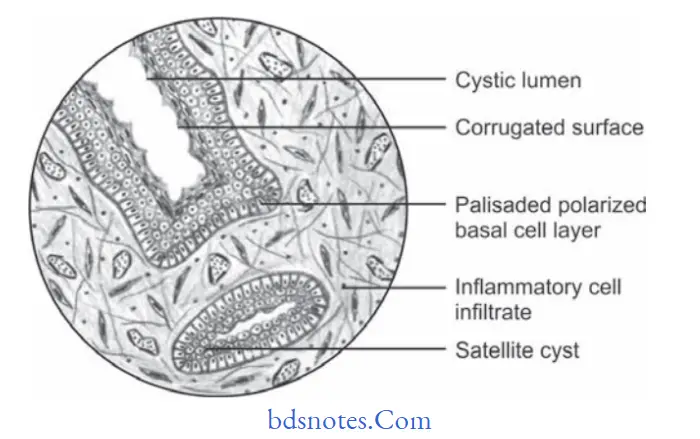
Histopathological Features of Odontogenic Keratocyst
- A parakeratin surface which is usually corrugated rippled or wrinkled.
- Uniformity of thickness of epithelium is generally between 6 to 10 cells in depth.
- Prominent palisaded, polarized basal cell layer often described as having a “picket fence” or “tombstone”appearance.
- Occasionally orthokeratin is found but if present, parakeratin is evident.
- Connective tissue shows “Daughter cells” or “Satellite cysts”.
- Lumen of keratocyst may be filed with thin straw-colored flid or with thick creamy material.
- Sometimes a lumen contains a great deal of keratin while at other times it has litte cholesterol as well as hyaline bodies at the site of inflmmation.
OKC jaw cyst
“Common challenges in diagnosing odontogenic keratocysts effectively: FAQs provided”
“Steps to explain causes of odontogenic keratocysts: Genetic mutations vs developmental factors: Q&A guide”
Clinical Features of Odontogenic Keratocyst
- Peak incidence is between 2nd and 3rd decades of life.
- It is found more frequently in males as compared to females.
- Mandible is affcted more commonly than maxilla.
- In mandible, the majority of cysts occurs in ramus third molar area, followed by fist and second molar area and then the anterior mandible.
- It is asymptomatic unless they become secondarily infected in which case patient complains of pain, soft tissue swelling and drainage.
- Occasionally, they experience paresthesia of lower lip and teeth.
- There is often one tooth missing from the dental arch.
- Expansion and thinning of bone may result in pathological fracture.
- Maxillary odontogenic keratocyst tends to be secondarily infected with greater frequency than the mandibular ones,due to its vicinity to maxillary sinus.
“Role of PTCH1 gene mutation in causing odontogenic keratocysts: Questions answered”
Radiological Features of Odontogenic Keratocyst
- Odontogenic keratocyst is oval in shape and it extends to the body of mandible with mediolateral expansion.
- It is very small in size or it can exceed the diameter of 5 cm.
- Margins of the cyst are hyperostotic.
- Mostly odontogenic keratocyst is unilocular and have smooth borders while some of the cysts show irregular borders too.
- Radiolucency is seen in the cystic part which appears to be hazy if keratin is present in the cavity.
- Radiolucency is surrounded by thin sclerotic rim.
- In some of the cases perforation of lingual and buccal cortical plates is seen.
- Displacement of inferior alveolar canal is seen downwards.
Leave a Reply