Odontogenic Cysts
Question. Write a short note on the radiographic appearance of periapical cyst.
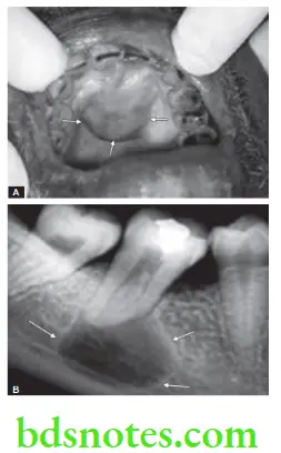
Answer. It appears as a rounded or pear-shaped radiolucency at the apex of the non-sensitive tooth or with the nonvital tooth.
- Radiolucency is more than 1.5 cm in diameter but usually less than 3 cm in diameter. It has got well-defined outline with thin hyperostotic borders.
- Margins: In uncomplicated cases, margins are smooth, corticated and cortex is usually well-defined, well-etched and continuous, except in some cases, there may be window formation. There is also thin white line surrounding the margins of bone cavity. This thin layer of cortical bone is almost always present unless suppuration supervenes in the cyst.
- Image of radiopaque borders is continuous with lamina dura around the associated tooth. Infection may cause the borders to become less distinct.
- Radicular cysts of long duration may cause resorption of roots.
Odontogenic Cysts: Types, Symptoms, and Treatment
- Adjacent teeth are usually displaced and rarely resorbed.
- If the maxillary area is involved, there is displacement of the maxillary sinus.
Leave a Reply