Occlusal Radiography
Write a short note on occlusal radiography.
or
Write a short note on occlusal radiographic technique.
or
Write a short note on occlusal X-ray.
or
Discuss occlusal radiographs in detail.
or
Write a short note on indications of occlusal radiography.
or
Write a short answer on indications of occlusal radiography.
or
Write a short answer on the occlusal radiograph.
Answer. It is an intraoral radiographic technique.
- This technique is used to examine large areas of the upper and lower jaw.
- The palate and floor of mouth may also be examined.
- This is a supplementary radiographic technique that is used in conjunction with periapical or bitewing radiography.
“Role of occlusal radiography in identifying impacted teeth and cysts: Questions answered”
Occlusal Radiograph Indications
- In locating, the retained roots of extracted teeth.
- In locating, the supernumerary, unerupted or impacted teeth (especially impacted canine and third molars).
- In locating, the foreign bodies in the maxilla or mandible.
- In locating, the salivary stones in the duct of the submandibular gland.
- In locating and evaluating the extent of lesions (e.g. cysts, tumors, malignancies) in the maxilla or mandible. It is especially indicated to determine the mesial and lateral extent of the lesion and its extent on the palate.
“Understanding the role of occlusal radiography in dental diagnostics: Q&A explained”
Types of occlusal radiographs
- In evaluating, the boundaries of the maxillary sinus (anterior, mesial and lateral outline).
- In evaluating, fractures of the maxilla and mandible. (location, extent and displacement).
- To aid in the examination of patient’s who cannot open their mouths more than a few millimeters or in adults and children who are unable to tolerate periapical films.
- To examine area of cleft palate.
- To measure changes in the size and shape of the maxilla and mandible.
- As a midline view, when using the parallel method for determining the buccal/palatal position of unerupted canines.
Maxillary occlusal radiograph
“Importance of studying occlusal radiography for better imaging outcomes: Questions explained”
Occlusal Radiograph Classification
- Classification
- Maxillary
- Cross sectional
- Topographic
- Anterior
- Posterior or lateral
- Pediatric
- Mandibular
- Cross sectional
- Topographic
- Anterior
- Posterior or lateral
- Pediatric
- Maxillary
Indications of occlusal radiograph
Occlusal Radiograph Basic Principles
- Film is positioned with white side facing the arch that is being exposed.
- Film is placed in mouth between occlusal surfaces of maxillary and mandibular teeth.
- Film is stabilized when the patient gently bites on the film surfaces.
- For maxilla: Upper arch is parallel to the floor and midsagittal plane is perpendicular to the floor.
- Formandible: The occlusal plane is perpendicular to the floor.
Cross-sectional occlusal radiograph
“Common challenges in performing occlusal radiography effectively: FAQs provided”
Occlusal Radiograph Film
- Occlusal films are bigger
- Ultraspeed films
- The films are coated on both sides
- Screen type is used in cassette and non-screened films are used in pocket
- For maxilla: Patient is asked to hold the cassette or packet with both the thumbs
Pediatric occlusal radiography
Various occlusal Radiographic Views and their techniques
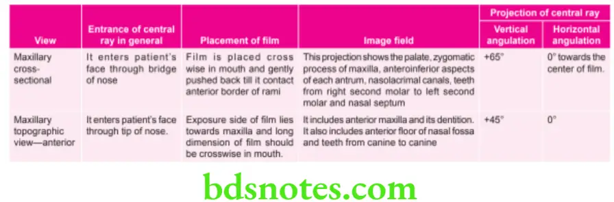
“Factors influencing success with occlusal radiography techniques: Q&A”

Maxillary Pediatric Occlusal Projection
- Here, the patient should be positioned upright with the maxillary arch parallel to the floor, so that sagittal plane is perpendicular to the floor and the occlusal plane is horizontal.
- Place a size–2 film with the white part facing the maxilla and the long edge over side to side-to-side direction.
Difference between occlusal and periapical
“Steps to apply occlusal radiography in diagnosing dental pathologies: Q&A guide”
- The film should be inserted in the child’s mouth, place the film as far posteriorly as the patient’s anatomy permits till it contacts the anterior border of the ramus of the mandible.
- The child should be asked to bite down gently on the film, retaining the position of the film in an end-to-end bite.
Uses of occlusal radiography
- Position indicating device (PID) is positioned so that the central ray is directed via the midline of the arch towards the centre of the film at a vertical angulation of +600. Top edge of the position indicating device is placed between the eyebrows on the bridge of the nose. In general, the central ray enters the face of the patient via the bridge of the nose.
Leave a Reply