Non-Odontogenic Connective Tissue Malignant Tumors: A Focus on Fibrosarcoma
Question. Enumerate non-odontogenic connective tissue malignant tumors. Discuss in detail about clinical features, etiology, and histopathology of fibrosarcoma.
Answer. Enumeration of non-odontogenic connective tissue malignant tumors
- Fibrous connective tissue: Fibrosarcoma
- Adipose Tissue: Liposarcoma
- Cartilage: Chondrosarcoma
- Bone
- Osteosarcoma
- Osteochondrosarcoma
- Vascular:
- Hemangioendothelioma
- Angiosarcoma
- Kaposi sarcoma
“Importance of studying fibrosarcoma for better diagnostic outcomes: Questions explained”
- Neural tissue: Neurosarcoma or Neurofirosarcoma
- Muscle
- Leiomyosarcoma
- Rhabdomyosarcoma
- Lymphoid tissue
- Hodgkin and NonHodgkin lymphoma
- Lymphosarcoma
- Reticular cell sarcoma
- Ewing’s sarcoma
- Burkitts lymphoma
- Multiple myeloma
- Leukemia.
“Understanding non-odontogenic connective tissue malignant tumors: Q&A explained”
Fibrosarcoma
- Fibrosarcoma is the malignant firous connective tissue tumor and is the malignant tumor of firoblasts.
Fibrosarcoma Clinical Features
- Fibrosarcoma arises at any age but mean age is 40 years.
- Male predilection is seen.
- Fibrosarcoma is most commonly seen in lower extremities i.e. femur and tibia.
- In oral cavity tumor involves mandible, maxilla, maxillary sinus, lip and palate.
- Tumor is generally a large painless mass which lies deep to fascia and has ill-defied margin.
- Associated teeth become mobile.
- Tumors in starting show benign growth and later on they spread rapidly producing large tumor with ulceration and hemorrhage.
- They can also cause pathological fracture.
“Common challenges in diagnosing fibrosarcoma effectively: FAQs provided”
Fibrosarcoma Etiology
- Most of the firosarcomas arise from preexisting lesions such as Paget’s disease, firous dysplasia chronic osteomyelitis, bone infarcts and in previously irradiated areas of bone.
- Congenital firosarcomas are thought to arise from genetic mutations.
Fibrosarcoma Histopathology
- Various histological grading of firosarcoma are:
“Steps to explain causes of fibrosarcoma: Genetic mutations vs environmental factors: Q&A guide”
“Role of connective tissue dysregulation in causing fibrosarcoma: Questions answered”
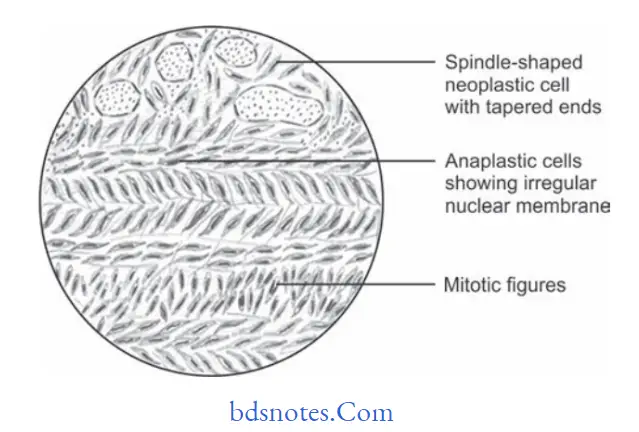
Fibrosarcoma Well-differentiated
- In this multiple plump-shaped firoblasts with pale eosinophilic cytoplasm, hyperchromatic spindleshaped nuclei with tapered ends is seen.
- Malignant firoblasts are dispersed in rich collagen area.
- Few mitotic fiures are evident.
“Early warning signs of issues addressed by understanding fibrosarcoma pathogenesis: Common questions”
Fibrosarcoma Intermediate Grade
- In this tumor show Herring bone pattrn, i.e. parallel sheets of cells arranged in intertwining whorls.
- Cellularity is high.
- Cellular pleomorphism is evident.
- Areas of hyalinization can be appreciated.
Fibrosarcoma High Grade
- Marked cellular atypia and mitotic activity is evident.
- This grade is highly anaplastic and pleomorphic with bizarre nuclei.
Leave a Reply