Nerves Of The Orbit
“What are the nerves of the orbit? A detailed question and answers guide”
Nerves of the orbit (or) Lateral rectus muscle of eyeball
Answer:
1. Lateral Rectus Muscle Of Eyeball Optic nerve:
- Lateral Rectus Muscle Of Eyeball Optic nerve is the nerve of sight
- Lateral Rectus Muscle Of Eyeball Optic nerve is made up of the axons of cells in the ganglionic layer of the retina
- Lateral Rectus Muscle Of Eyeball Optic nerve emerges from the eyeball 3 or 4 mm nasal to its posterior pole
- Lateral Rectus Muscle Of Eyeball Optic nerve is enclosed in three meningeal sheaths
- Lateral Rectus Muscle Of Eyeball Optic nerve cannot degenerate once it is cut
2. Lateral Rectus Muscle Of Eyeball Oculomotor nerve:
- Lateral Rectus Muscle Of Eyeball Oculomotor nerve is third cranial nerve
- Lateral Rectus Muscle Of Eyeball Oculomotor nerve is distributed to the extraocular & intraocular muscles
- Functional components:
- General somatic efferent
- General visceral efferent
- General somatic afferent
“Understanding the nerves of the orbit through FAQs: Composition, functions, and uses explained”
Optic nerve Nucleus:
- Optic nerve Nucleus is situated in the ventromedial part of central grey matter of midbrain at the level of superior colliculus
3. Trochlear nerve:
- Trochlear nerve is fourth cranial nerve
- Trochlear nerve supplies superior oblique muscle of the eyeball
- Functional components:
- General somatic efferent
- General somatic afferent
“Importance of studying nerves of the orbit for medical students: Questions explained”
Nucleus:
- Nucleus is situated in the ventromedial part of central grey matter of midbrain at the level of inferior colliculus
4. Abducent nerve:
- Abducent nerve is sixth cranial nerve
- Abducent nerve supplies the lateral rectus muscle of the eyeball
- Abducent nerve Functional components:
- General somatic efferent
- General somatic afferent
Abducent nerve Nucleus:
Abducent nerve Nucleus is situated in the upper part of the floor of fourth ventricle in the lower pons
5. Branches of ophthalmic & maxillary divisions of the trigeminal nerve:
- Branches of ophthalmic divisions:

“Common challenges in mastering nerves of the orbit notes effectively: FAQs provided”
- Branches of maxillary division:
- Infraorbital nerve:
- Infraorbital nerve enters the orbit through the inferior orbital fissure
Branches:- Middle superior alveolar nerve
- Anterior superior alveolar nerve
- Terminal branchespalpebral, nasal & labial
- Infraorbital nerve enters the orbit through the inferior orbital fissure
Abducent nerve orbit course
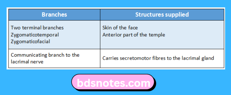
- Zygomatic nerve:
- Zygomatic nerve enters the orbit through the lateral end of the inferior orbital fissure
Branches:
“Factors influencing success with nerves of the orbit studies: Q&A”
6. Sympathetic nerves:
- Arises from the internal carotid plexus
- Sympathetic nerves enter the orbit through
- Ophthalmic nerve
- Nasociliary nerve
- Long Ciliary branches
- Plexus surrounding ophthalmic artery
- Direct branch from internal carotid plexus
- Oculomotor, Trochlear, Abducent & ophthalmic nerves
Leave a Reply