Neisseria
Question 1. Write a short note on gonococci.
Answer:
Gonococci lead to sexually transmitted diseases known as gonorrhea.
Morphology
- They are gram-negative cocci.
- The size of gonococci is 0.6 to 1 µm in diameter.
- Gonococci are either pear or bean-shaped, and it possess pili on its surface.
- They are arranged in pairs with concave adjacent sides.
- They are non-motile as well as non-sporing.
- Gonococci are capsulate
Read And Learn More: Microbiology Question And Answers
“Understanding Neisseria through FAQs: Q&A explained”
Gonococci Cultural Characteristics
- Gonococci can be grown aerobically or anaerobically.
- They grow at a temperature of 35-36°C in the presence of 5–10% carbon dioxide and at pH is 7.2-7.6.
- They require an enriched medium chocolate agar for their growth.
- They can also be grown on a selective medium, i.e. Thayer Martyn medium which consists of chocolate agar with antibiotics.
Gonococci Colonies
- Small, round, convex or slightly umbonate, translucent, grey with finely granular surface.
- According to their colony appearance, autoagglutinability, and virulence, Kellogg divides colonies of gonococci into two types, which are as follows:
- Type T1 and Type T2: These are small brown colonies that are produced by piliated strains, autoagglutinable and virulent gonococci.
- Type T3 and Type T4: They are produced by non-piliated avirulent strains which form smooth suspension.
- Type T1 and T2 are produced by fresh isolates and on sub-culture they become T3 and T4 colonies.
“Importance of studying Neisseria for microbiology students: Questions explained”
Gonococci Biochemical Reactions
- It is catalase positive
- It is oxidase positive
- It utilizes glucose only with the production of acid.
Antigens Present in Gonococci
- Pili protein: By recognizing the specific receptors it helps in attachment with host cells. It inhibits phagocytosis too.
- Outer membrane protein I, II, III: Protein I, i.e. por protein, and Protein III, i.e. Rmp protein, both act as ligands and attach gonococci to host cells. Protein I causes formation of pores and protein III is associated with it. Protein II, i.e. Opa protein is associated with the adherence of gonococci to host cells and also for the clumping of cocci in urethral exudates. Protein II also inhibits phagocytosis.
- IgA protease: It causes cleavage of IgA and inactivates it.
- Lipopolysaccharide: It produces endotoxic effects so there is toxicity of infection.
- Capsule: Seen in freshly isolated strains. It is polyphosphates that allow the intracellular survival of the organism
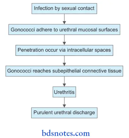
Pathogenesis
“Common challenges in understanding Neisseria effectively: FAQs provided”
“Factors influencing success with Neisseria knowledge: Q&A”
Question 2. Discuss the laboratory diagnosis of gonorrhea.
Answer:
Gonorrhea is a bacterial disease caused by Neisseria gonorrhoeae.
Specimen collected is urethral discharge and cervical discharge:
1. Direct Microscopy:
- Gram staining of the smear show gram-negative intracellular diplococci and plenty of pus cells.
- Fluorescent antibody technique: In this smear is prepared and is treated with an antibody conjugated to fluorescent tag—showing the presence of bacteria. It is more specific.
“Role of pili and capsules in Neisseria pathogenicity: Questions answered”
2. Detection of Antibody:
This is done in patients having metastatic lesions, especially when isolation falls. Tests used are ELISA, radioimmunoassay, and hemagglutination.
3. Culture:
Chocolate agar or blood agar media is used for the culture of organisms and these are incubated at 35°C in 5 to 10% carbon dioxide for 48 hours. Translucent colonies appear over the culture medium. Colony morphology is seen which reveals small, round, gray, translucent, and convex colonies with fie granular surfaces. Selective mediums such as Thayer-Martin can also be used for all genital rectal samples.
4. Superoxol Test: N. gonorrhea produces brisk bubbling when colonies are emulsified by 30% hydrogen peroxide on a glass slide.
5. Compliment Fixation Test: It becomes positive in some weeks as the infection is established and remains positive for months and years as the disease becomes cured.
“Steps to explain types of Neisseria: Gonorrhea vs meningitis pathogens: Q&A guide”
6. Biochemical Tests:
- It is catalase positive
- It is oxidase positive
- It utilizes glucose only with the production of acid.
- It does not ferment maltose
7. Antibiotic susceptibility testing: By Kirby – Bauer disc diffusion method on gonococcus agar with growth supplements using ceftriaxone, ciproflxacin, ofloxacin, tetracycline, doxycycline, erythromycin, etc. Gonococci develop resistance to penicillin.
Leave a Reply